Nausea, vomiting, left upper quadrant pain: A POCUS case of gastric volvulus
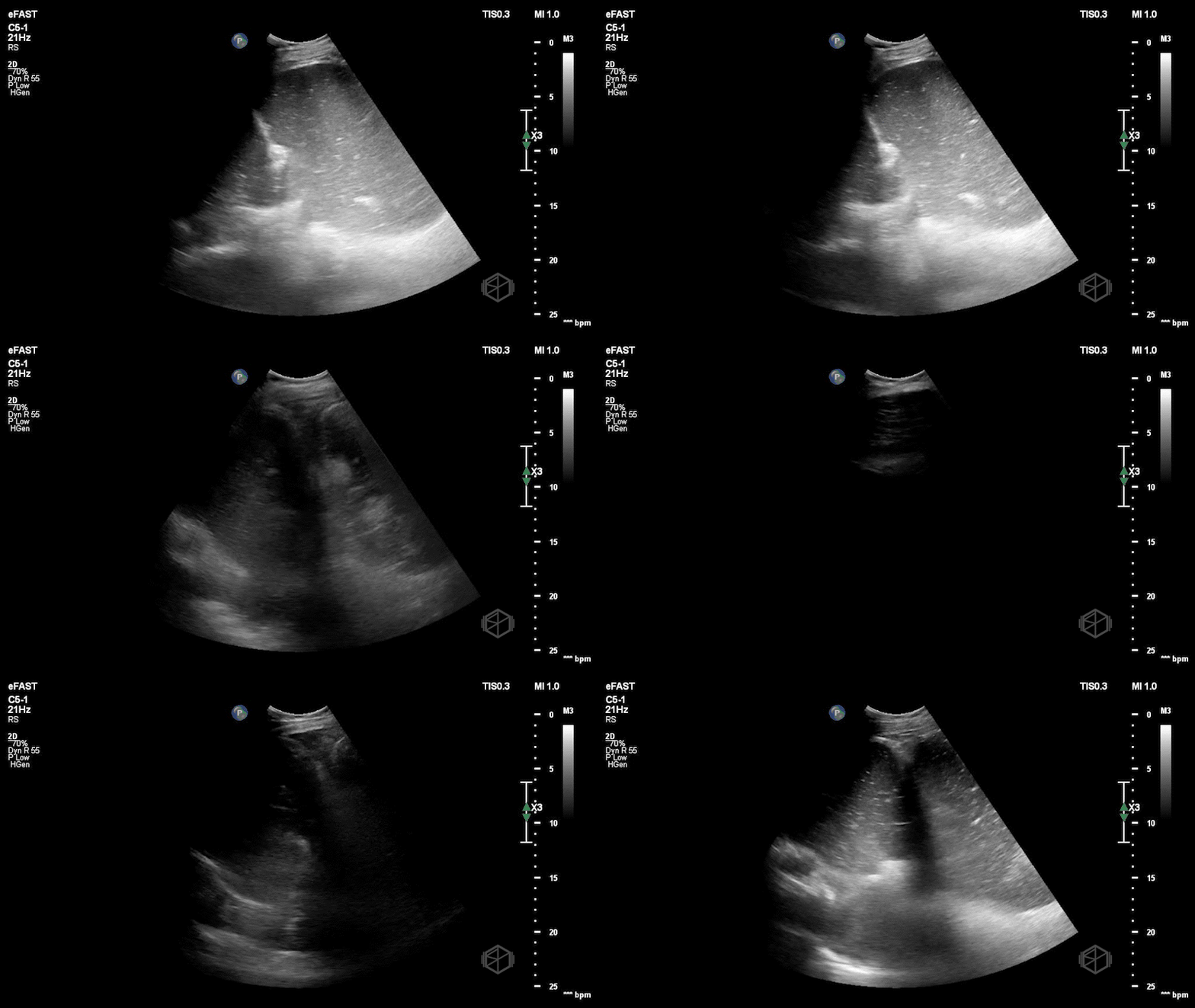
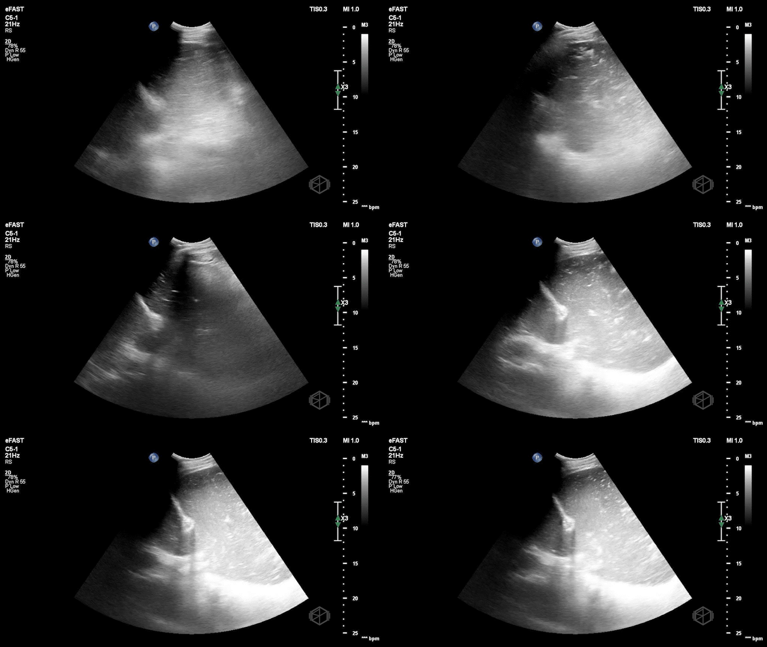
Left upper quadrant ultrasound demonstrating a hiatal hernia with stomach herniating through.
The stomach is swirling with food contents and is visible on both sides of the diaphragm.
CT scan showing marked distention of the stomach with a large hiatal hernia and gastric volvulus with obstruction of the gastroduodenal junction and lesser obstruction at the gastric body most likely representing an organoaxial gastric volvulus.
May Ali, DO
PGY-1, Emergency Medicine
Yash Chavda, DO, MBA, FAAEM
Clinical Assistant Professor
NYU Grossman Long Island School of Medicine
Director of Emergency Ultrasound
NYU Langone Hospital — Long Island
Christopher Caspers, MD, MBA
Clinical Professor
NYU Grossman Long Island School of Medicine
Chair, Department of Emergency Medicine
NYU Langone Hospital — Long Island
References
Hassan A, Azhar A, Ullah A, et al. Gastric organoaxial volvulus: A lethal twist and a rare cause of acute abdomen. Radiol Case Rep. 2023;18(11):4076-4079. doi:10.1016/j.radcr.2023.08.012
Jakubowski J, Lizzi J, Hill T. Gastric Volvulus. Journal of Education and Teaching in Emergency Medicine. Published online July 1, 2019. doi:10.21980/J8335F
Matsuzaki Y, Asai M, Okura T, Tamura R. Ultrasonography of gastric volvulus: “peanut sign.” Intern Med. 2001;40(1):23-27. doi:10.2169/internalmedicine.40.23
Shokraneh K, Johnson J, Cabrera G, Kalivoda EJ. Emergency Physician-Performed Bedside Ultrasound of Gastric Volvulus. Cureus. 12(8):e9946. doi:10.7759/cureus.9946


