December SonoProps
This month was another incredibly tough month of scans with a lot of high quality scans and interesting pathology.
However, December brings many surprises, so this month’s theme is surprises…
The first SonoProps goes to our current resident rotator Dr. Antonia Nevias-Ida.
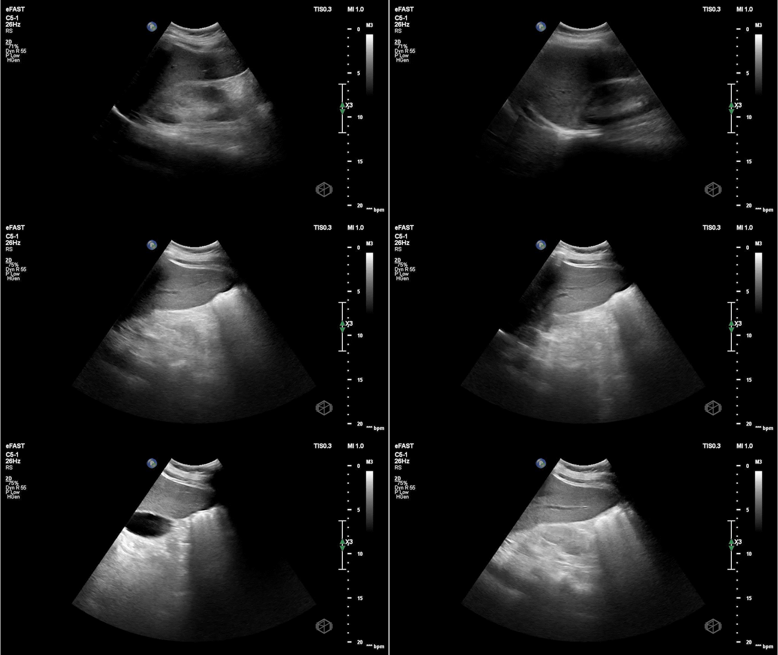
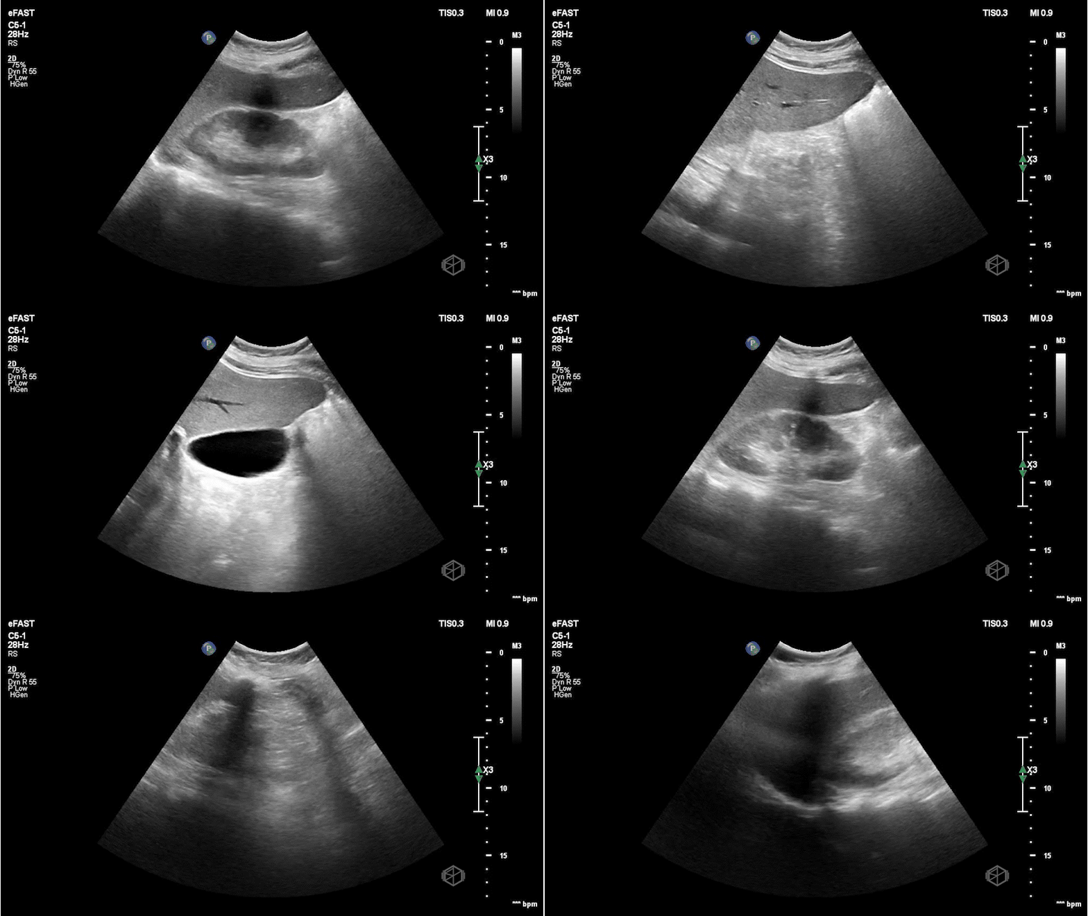
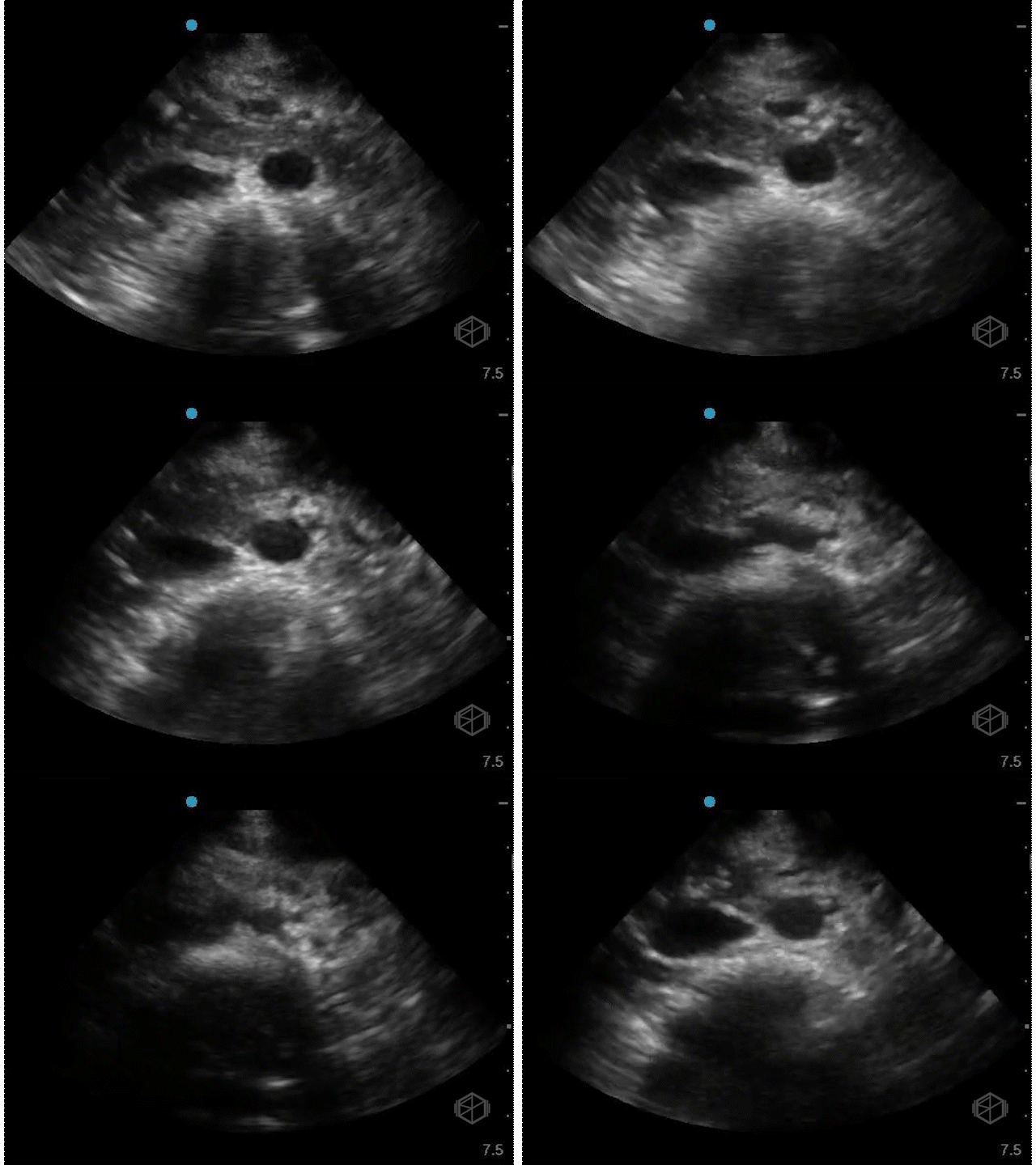
She was scanning a 55-year-old female with LEFT flank pain that came on sudden onset and captured the following images:
If you are thinking this is positive for free fluid — surprise — it is not!
It is actually an artifact called lipliner sign. Free fluid generally makes sharp angles and persists throughout multiple views, where as this anechoic stripe disappears as the probe position is varied.
Diagnosis: Lipliner sign
Learning points:
There are many misleading artifacts and false positives to consider when performing a FAST/FAFF examination including focal fatty sparing, double line sign, and now the lipliner sign.
Lipliner sign is an artifact that appears at the edge of solid organs such as the liver or the spleen (the more common areas for free fluid) (39366788) and can easily mislead a clinician to assume there is free fluid present.
It is not definitively known what causes the lipliner sign but it may be due to advances in ultrasound machines likely due to adaptive filtering to improve spatial resolution and post-processing technology.
The artifact may be visible on machines from multiple manufacturers.
When encountering lipliner artifact and deciding it may be reasonable to call the FAST equivocal rather than a true positive if there is any question.
True free fluid makes sharp angles, dissects through structures, is dependent, should not have any membranes or walls, and is dynamic (eg, place the patient in Trendelenburg and more fluid will shift up).
The next SonoProp goes to Dr. Obioma Nkemakolam.
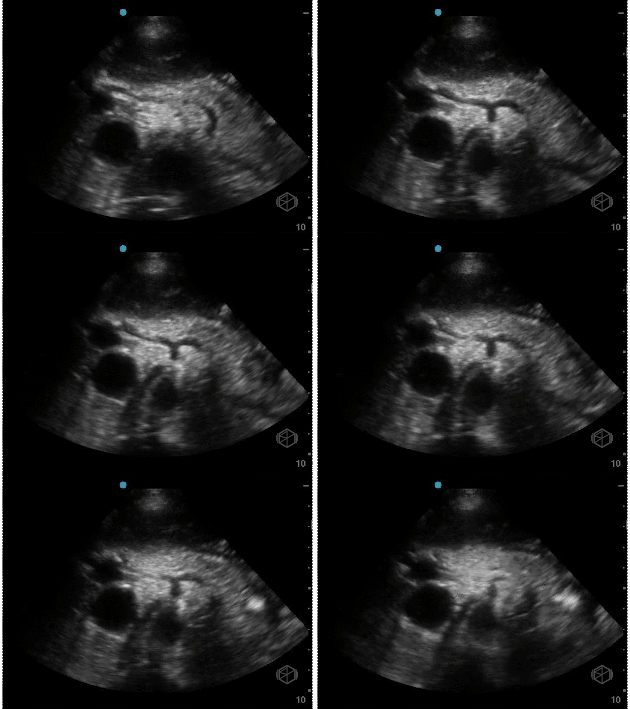
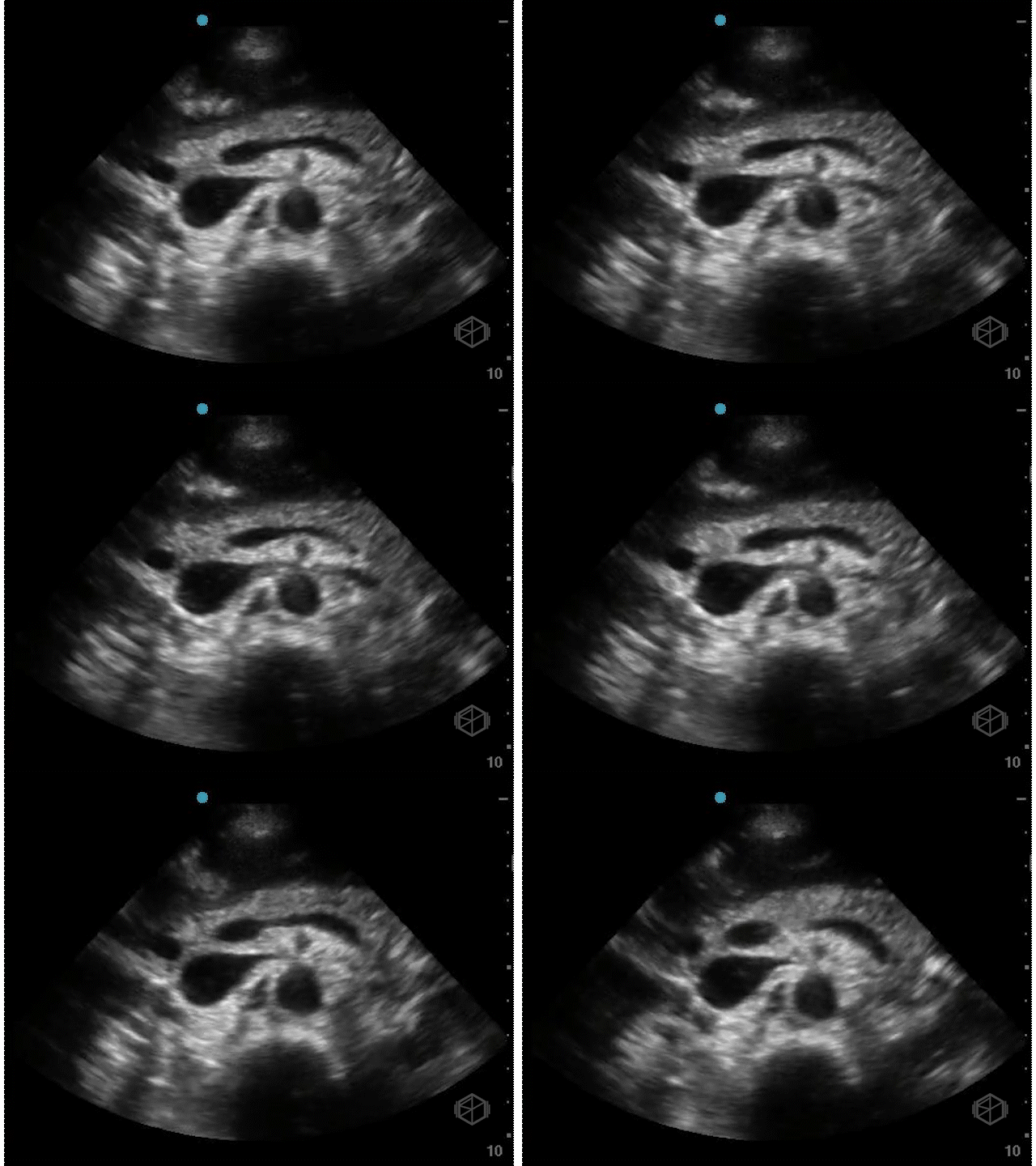
He was evaluating a 54-year-old male with a history of hypertension, hyperlipidemia, coronary artery disease, and current everyday smoking, who presented with shortness of breath. The patient was significantly hyperglycemic and was thought to be in DKA. He was also borderline hypotensive and there was a lacy appearance to his abdomen. The patient had a stat abdominal aorta scan that demonstrated the following:


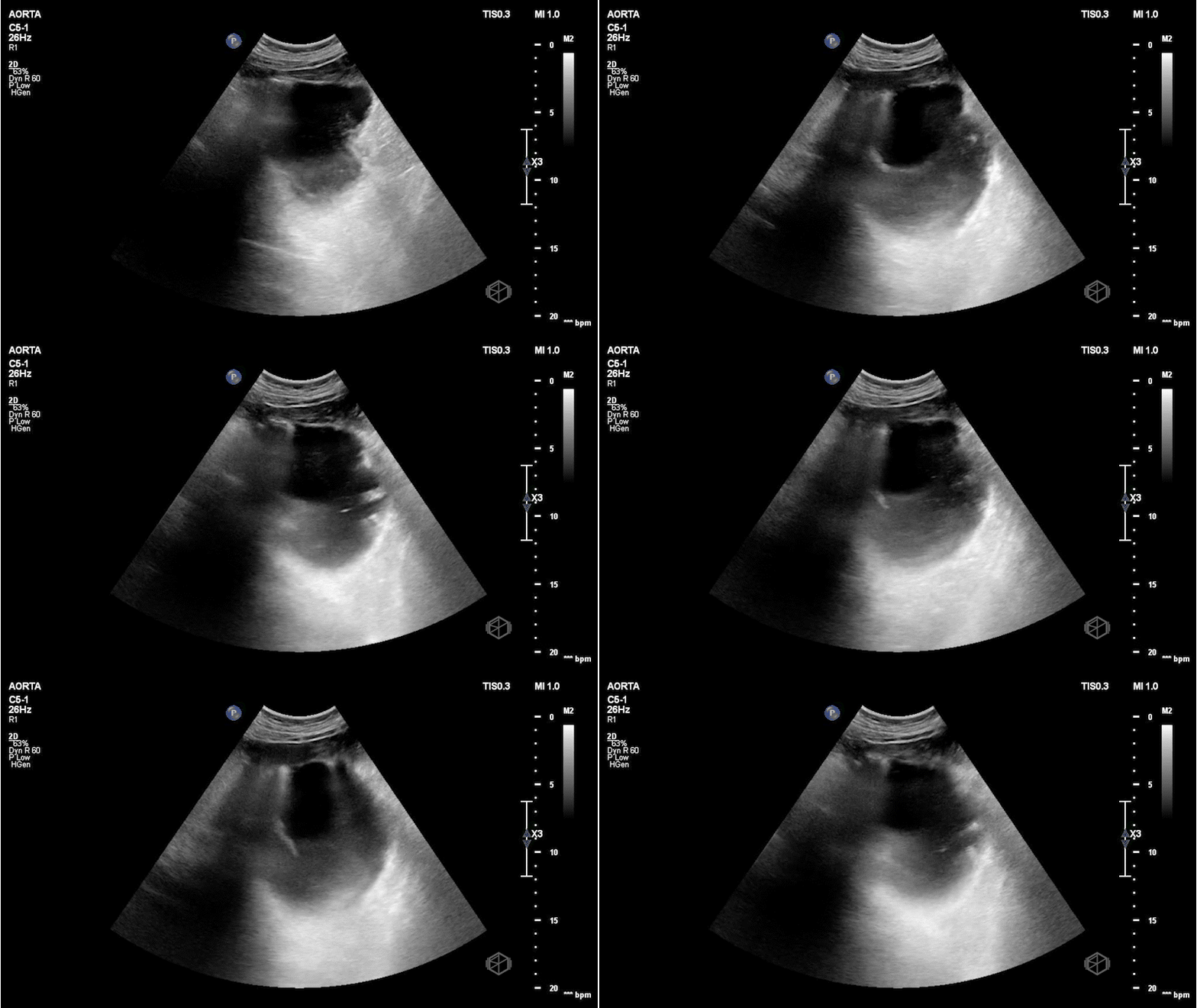
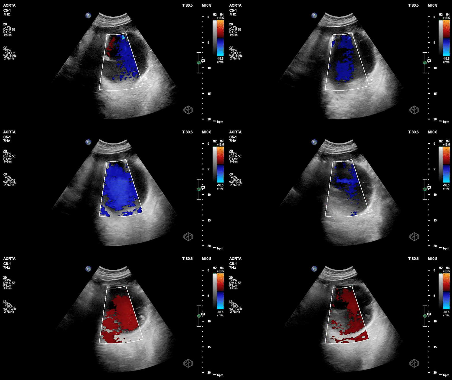
There is a large circular anechoic structure in the middle of the scan. If you thought this was the abdominal aortic aneurysm — surprise — it’s not.
This a distended fluid filled stomach, a common false positive for FAST/FAFF examinations as well as aorta exams.
Patient had a CTA that demonstrated a normal aorta as well as diffuse gaseous distention of the esophagus mild/moderate gastric distention with fluid and gas due to recent alimentation.
Diagnosis: Distended stomach filled with liquid/food
Learning points:
The aorta has some key landmarks that should be identified before determining the structure is the aorta on ultrasound - the vertebral body should be right behind it. The IVC should be next to it on the left. The spinal body should be visualized. The aorta is also pulsatile and will have the celiac trunk (if the proximal aorta can be visualized) and superior mesenteric artery visible.
This structure seems to be coming from the left upper quadrant, with no vertebral body behind it and no IVC next to it. The color doppler signal is unreliable due to the patient’s heavy breathing.
The stomach will appear as a cystic organ when distended with fluid. If there is clear fluid there may be echogenic gas bubbles visible (28191199). There many hints that the object in Dr. Nkemakolam’s case is the stomach and not the aorta.
Breathing and peristalsis of the stomach may cause doppler noise.
Diagnosis of stomach pathologies (eg, malignancy, Menetrier’s disease, hiatal hernia, gastric volvulus) may be possible with stomach ultrasonography although this is highly operator dependent and patient dependent and is usually not performed as dedicated emergency point-of-care ultrasound (23329910). There is anesthesia literature regarding use of stomach ultrasonography for perioperative planning.

