
January 2026 SonoProps
🎉 Happy New Year, Everyone!
To kick off the new year, we’re highlighting SonoProps cases where POCUS revealed a surprise finding and meaningfully changed patient care.
🏆 SonoProp #1 – Dr. Hardeep Singh
Dr. Singh had a 20’s male with a past medical history of HTN, LVH, congenital bicuspid aortic valve, obesity, HLD, single coronary artery system present to the ED with chest pain and near syncope.
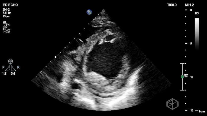
Dr. Singh grabbed the US and saw the following findings:
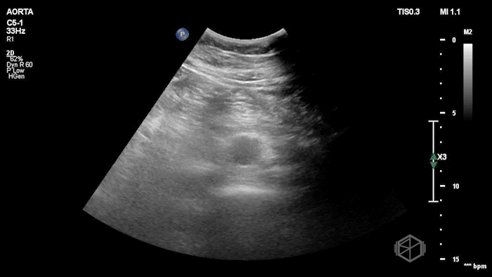
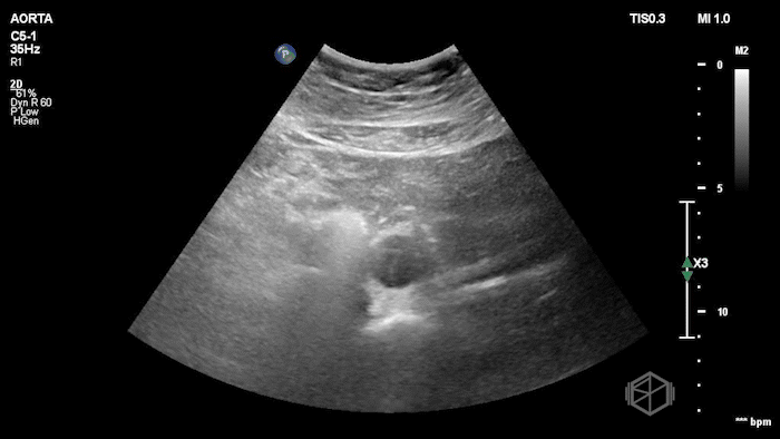
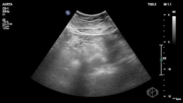
The POCUS shows a normal sized abdominal aorta, but with a visible flap moving internally. Given the patient has chest pain and near syncope, and extensive dissection was in the differential. Dr. Singh and Dr. Bernot expedited the patient to CTA which showed an extensive Type A aortic dissection. The patient went immediately to surgery and had a positive outcome.
Diagnosis: Type A Aortic Dissection
Learning Points:
POCUS Can Detect an Intimal Flap, Highly Specific
Multiple case reports show that visualizing a mobile intimal flap on POCUS (in the abdominal aorta or cardiac views) is a specific sign of aortic dissection. It’s not common, but when seen it definitely get the definitive test expedite the patient’s care. (📚 PMID: 30443611)
POCUS Is Not Sensitive Enough to Rule Out Aortic Dissection
A systematic review of emergency physician–performed POCUS for thoracic aortic dissection found a wide range of sensitivities (≈41–91%) when an intimal flap is seen — but specificities are high (≈94–100%). This pattern means POCUS is specific when positive but not reliable as a stand-alone rule-out tool (📚 PMID: 37364927).
Structured POCUS Protocols Have Strong Diagnostic Performance
The SPEED protocol — which combines abdominal aorta views, cardiac assessment — showed a sensitivity of ~93% and specificity ~91% for acute aortic dissection in one prospective observational study (N ≈ 1314 exams) (📚 PMID: 38010071).
The protocol includes:
Cardiac POCUS
Assessment for pericardial effusion/tamponade
Evaluation of aortic root dilation
Identification of proximal intimal flap
Signs of acute aortic regurgitation
Abdominal aortic POCUS
Measurement of aortic diameter
Evaluation for intimal flap or true/false lumen
Integration with clinical suspicion, rather than use as a stand-alone rule-out test
POCUS Can Expedite Care in Unstable Patients
Case reports and series (e.g., detection of a pericardial effusion + intimal flap before CT diagnosis) illustrate POCUS can identify direct signs before advanced imaging — especially in clinical situations where transport to CTA is delayed or unsafe. (📚 PMID: 36299499)
While CTA remains the most common first-line definitive imaging modality for suspected aortic dissection; TEE offers comparable diagnostic accuracy and is particularly valuable in unstable patients who cannot safely leave the resuscitation bay. (📚 PMID: 8416265, 8529244, 36322642)
🏆 SonoProp #2 – Dr. Daniel Herzog
Dr. Herzog was working at NYU Suffolk where he had a mid 60’s female with reportedly only a past medical history of anxiety (recently diagnosed) presenting to the ED with anxious sensation, triaged for anxiety. The patient was slightly tachycardic and mild respiratory distress. Dr. Herzog grabbed the US and saw the following:
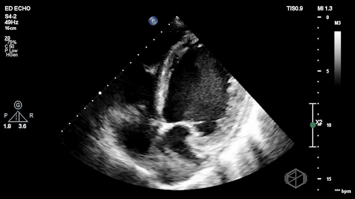
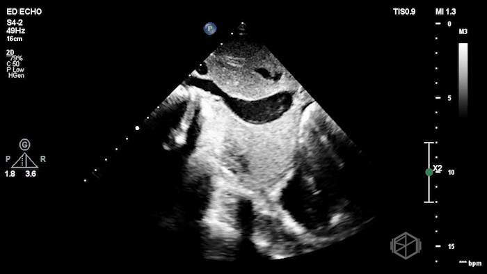
The POCUS shows an extremely low EF, about 15-20% visually, with a plethoric IVC. The patient had a cath that showed mild non-obstructive coronary disease and she was determined to have non-ischemic cardiomyopathy due to unknown etiology, possibly viral infection vs. stress related.
Diagnosis: New onset congestive heart failure with significantly reduced ejection fraction.
Learning Points:
Visual Estimation of LVEF by POCUS Is Accurate and Clinically Reliable
Multiple studies demonstrate that visual (“eyeball”) estimation of LVEF by trained emergency physicians correlates well with cardiology echocardiography. While not precise to single-digit percentages, visual estimation reliably categorizes EF as normal, moderately reduced, or severely reduced, which is often all that is needed for ED decision-making (📚 PMID: 11874773, 22044429, 24672616, 34654586)
“Anxiety” Should Be a Diagnosis of Exclusion, Especially in Older Patients with Dyspnea/Tachycardia
This case is a classic reminder that a label of anxiety can create anchoring bias. In an older patient with tachycardia and respiratory distress, cardiopulmonary causes must be evaluated first, and guideline-based ED risk stratification for chest pain equivalents prioritizes ruling out serious pathology. POCUS is uniquely positioned to break anchoring early by identifying dangerous physiology at bedside (📚 PMID: 34709879)
New Severe LV Dysfunction → Ischemic vs. Non-ischemic
When ED POCUS reveals new severe LV systolic dysfunction, patients should enter a pathway that considers ischemic heart disease early, because identifying CAD changes management. Guidelines recommend consideration of an ischemic evaluation in new-onset HF/newly reduced EF, commonly via coronary angiography or coronary CT, depending on clinical context. If obstructive CAD is excluded, the workup shifts toward non-ischemic cardiomyopathy etiologies (e.g., myocarditis, stress-induced cardiomyopathy, tachycardia-mediated, toxic/metabolic). (📚 PMID: 35363499, 40085773, 36328647)