July SonoProps
This month’s SonoProps were hard to decide. So many good scans. But, in the interest of variety and education the tough choice was made.
The first SonoProps goes to Dr. Marina Frayberg!
Dr. Frayberg had an approximately 80-year-old patient with recent URI type symptoms prescribed Azithromycin and Augmentin presenting to the ED with fevers and right knee swelling and pain. Due to this pain she could not ambulate.
A POCUS of the joint was performed:
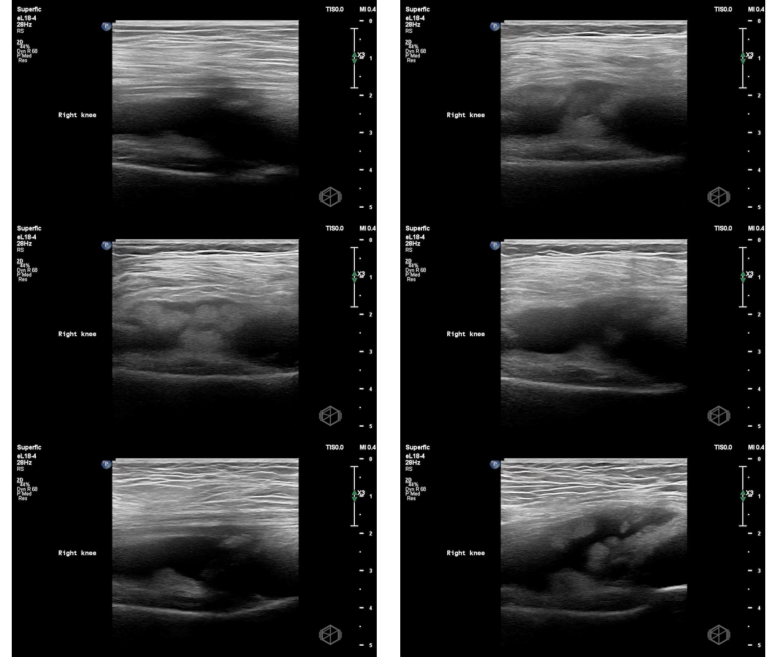
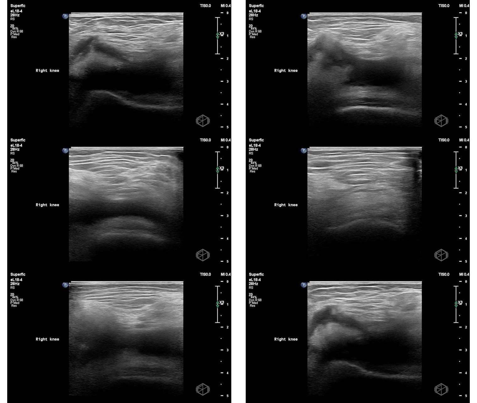
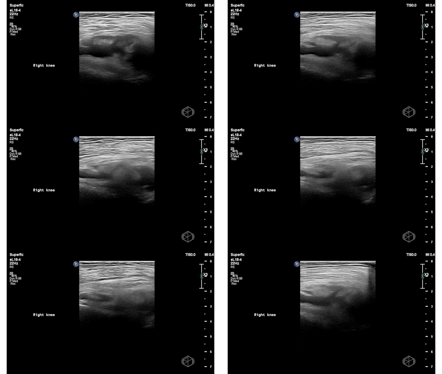
POCUS demonstrated a large, complex suprapatellar joint effusion.
Dr. Frayberg performed an ultrasound-guided arthrocentesis, successfully aspirating yellow fluid. Analysis revealed calcium pyrophosphate crystals. Gram stain and joint infection panel were negative.
Diagnosis: Pseudogout with large joint effusion
Learning points:
Probe & Position: Linear probe, patient supine, knee slightly flexed. Place probe longitudinally above the patella, probe marker toward the head. Then obtain transverse views (probe marker toward the right).
A simple effusion = anechoic (black), compressible, filling the suprapatellar recess.
A complex effusion = internal echoes, septations, or debris (suggests infection, crystals, or blood).
The differential diagnosis for a knee effusion includes:
Septic arthritis – purulent, echogenic fluid with debris and possible loculations; patient often febrile and joint significantly tender and hot (📚 PMID: 9231182).
Septic bursitis – similar findings but localized to a bursa, so the effusion is extra-articular (e.g., suprapatellar, prepatellar).
Crystal arthropathy
Gout: turbid synovial fluid due to monosodium urate crystals, may see echogenic foci or tophi (📚 PMID: 26995154).
Pseudogout: calcium pyrophosphate crystals; may appear cloudy or contain echogenic foci.
Hemarthrosis – blood in joint; can follow trauma or anticoagulation; often echogenic swirling fluid/clots.
Inflammatory arthritis (RA, psoriatic arthritis, etc.) – complex effusions from synovial proliferation and pannus formation.
Malignancy-associated effusion – rare; could be from primary synovial tumors (eg, synovial sarcoma) or metastatic disease, consider in patients with cancer history or persistent effusion without clear cause (📚PMID: 8587092).
Post-traumatic or post-surgical changes – fibrin strands, debris, and blood products may cause heterogeneity.
Ultrasound guidance for knee arthrocentesis improves first-pass success, increases fluid yield, and reduces pain—especially critical for novices and low-volume effusions, with less pain for the patient and shorter procedure times. (📚 PMID: 26791571, 35722054, 19062223).
The next SonoProps goes to Dr. Mary Jo Johnstone!
Dr. Johnstone had an approximately 50-year-old male presenting to the ED with right eye gradual vision loss x 6 days associated with floaters. Instead of calling a stroke alert, Dr. Johnstone grabbed the ultrasound which showed:
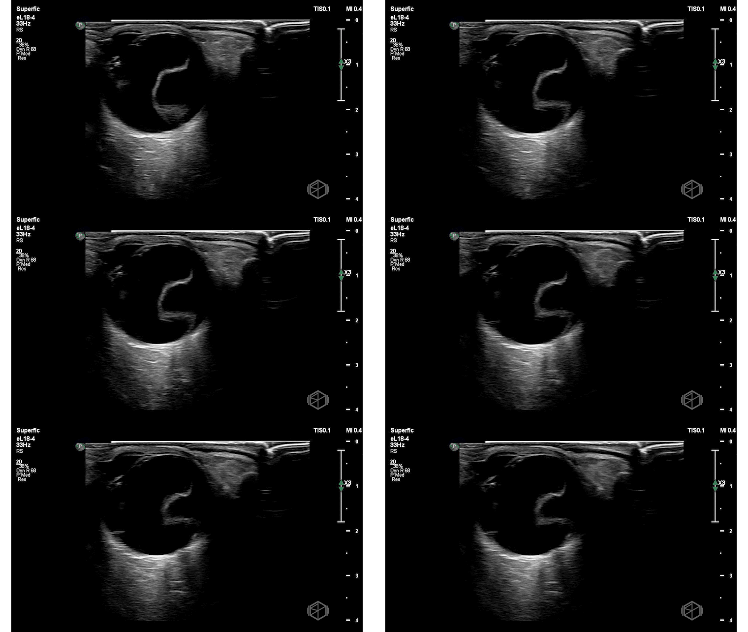
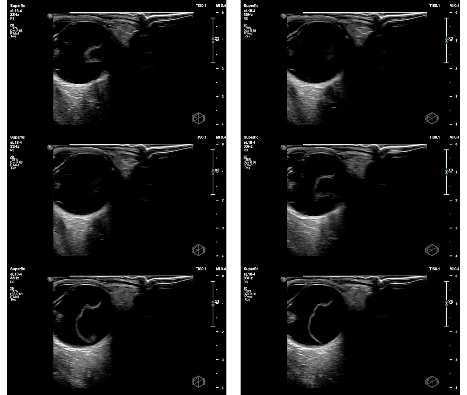
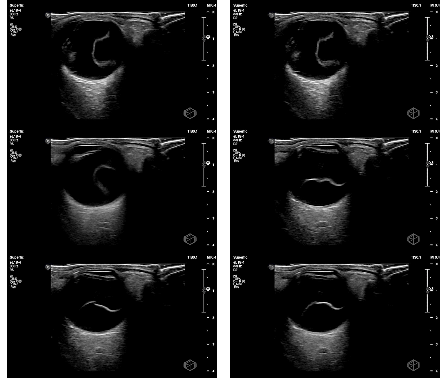
This ultrasound shows a hyperechoic linear flap attached the optic nerve. There is limited, taut “undulating” movement with oculokinetics.
Diagnosis: Right eye retinal detachment
Learning Points:
On B-scan ocular ultrasound, retinal detachment appears as a thick, hyperechoic membrane lifted off the posterior surface of the globe that floats and moves with the patient’s eye movements, especially when imaged with normal or low gain settings.
Large detachments are tethered to the optic nerve, while smaller ones remain closely attached to the posterior wall and show minimal movement. To improve detection of small retinal detachments, it is important to scan the eye in multiple planes, including transverse and sagittal axes.
Bedside ocular ultrasound by emergency physicians shows high sensitivity (97–100%) and moderate-to-high specificity (83–92%) for retinal detachment, making it a valuable rapid screening tool in the ED (📚 PMID: 20836770, 19625159, 12153883).
Another study showed that in a heterogenous group of emergency physicians - there was a high specificity (94%) but only moderate sensitivity (75%) for retinal detachment after minimal training. A positive scan strongly suggests RD, but a negative scan is not reliable to rule it out based on this study. Ophthalmology referral remains essential for patients with flashes or floaters, especially when novice or heterogenous operators are performing the POCUS (📚 PMID: 29774966). It is a rule in test, and not as good as a rule out test.
Determining Mac-ON vs. Mac-OFF is a more difficult task, but with experience and training it can be done.
If POCUS shows the the retina still attached to the macula (attachment point will be more lateral to the optic nerve) → “Mac-on” = TRUE ophthalmologic emergency. Central vision is preserved. Emergent repair needed to prevent progression and permanent central vision loss.
