June SonoProps
This month’s first SonoProps goes to Dr. Yanal Maher.
Dr. Maher was evaluating a 55-year-old female with an extremely distended young abdomen and abdominal discomfort. She reported that she had no prior medical problems but had not seen a doctor in a long time. The patient denied drinking alcohol. Physical exam demonstrated an extremely large protuberant abdomen that was firm to touch. No obvious spider angiomata were noted or significant skin changes.
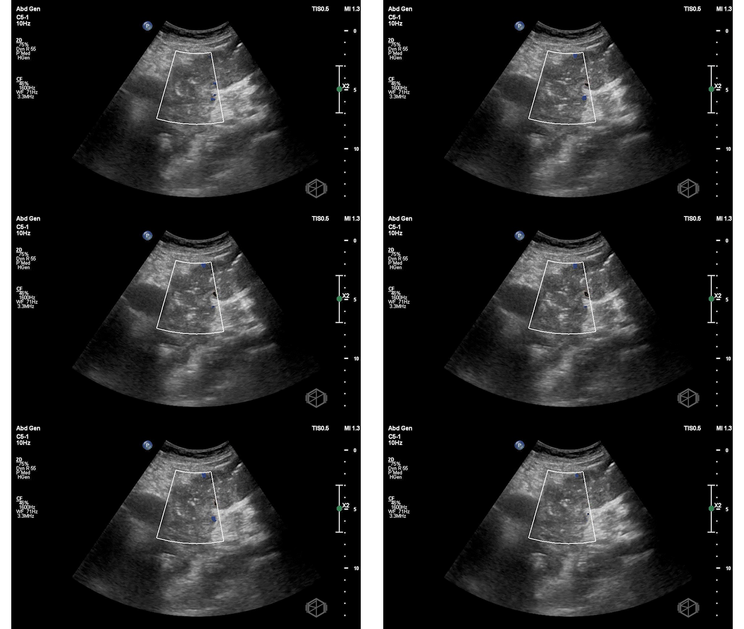
He performed a focused assessment for free fluid exam that demonstrated the following:
Upon placing the probe on the patients expected area of the RUQ, no obvious anatomical structures were noted (such as the kidney and liver). Instead, complex fluid was noted with internal debris appearing to be a mucous-like consistency.
Moving lower, there was a complex appearing structure that did not look like intestines as would be expected with ascites. There were septations noted within the structure. Moving higher up and significantly more posterior than usual, eventually the kidneys and liver were localized but significantly higher than expected. There did not appear to be any significant free fluid near the hepatorenal space.
The patient had a CT scan performed a while later that demonstrated a massive partially solid and cystic left adnexal lesion replacing the majority of the abdomen and pelvis with marked abdominal distention. This lesion measures at least 40.0 x 30 x 28 cm with a solid component anteriorly and superiorly measuring 18.0 x 10.8 x 9 cm. This mass also was compressing on the abdominal and pelvic vasculature although the patient had preserved flow.
Diagnosis: Large cystic mass likely of ovarian origin (eg, mucinous cystadenoma, cystadenocarcinoma etc.)
Learning Points:
It is important not to anchor on ascites—a distended abdomen is not always fluid. Think anatomically, and use imaging to differentiate.
Ultrasound findings of complex fluid with internal debris and septations suggest cystic structures rather than simple free fluid such as ascites. Simple ascites tracks to dependent areas—pelvis, paracolic gutters, and the hepatorenal recess—and typically reveals free-floating bowel and outlined organs. In this case, the intestines were not visible, and there were no classic fluid pockets. Instead, complex echogenic material with septations was noted.
(📚 PMID: 25493127)
Displacement of normal anatomy, such as the kidneys, liver, or intestines, can be a clue to the presence of a large mass rather than free fluid. In this case, the right upper quadrant was unrecognizable. Normal landmarks were displaced superiorly, suggesting a large intra-abdominal structure compressing surrounding organs. If you can’t find the anatomy where expected, it may not be a missed view—consider mass effect.
A firm, protuberant abdomen may indicate a mass rather than simple fluid accumulation, especially when percussion is dull and there is no shifting dullness. Tense ascites can present similarly, but the absence of perihepatic or perisplenic fluid and the lack of bowel floating in fluid on ultrasound helps differentiate the two.
Ovarian masses can be silent and reach massive sizes before detection, emphasizing the importance of routine health screening in women. (📚 PMID: 31196215)
Pseudomyxoma peritonei may occur with mucinous tumors and is a clinical syndrome caused by the accumulation of mucinous ascites due to rupture or leakage of mucin-producing tumors—most commonly from appendiceal neoplasms but also from ovarian mucinous tumors. The fluid typically appears mucinous and tracks along fascial planes. It may be visible around the liver and other organs, rather than displacing them as a large mass would. (📚 PMID: 36990788)
The second SonoProps of this month goes to Dr. Obioma Nkemakolam.
Dr. Nkemakolam evaluated an approximately 40-year-old patient who presented to the ED with intermittent bloody bowel movements for several months being worked up outpatient for inflammatory bowel disease. She had diffuse abdominal tenderness on exam. He performed a POCUS that showed the following:
Significant edematous bowel wall was noted extending from the rectum to the descending colon, transverse colon, and ascending colon indicating significant inflammation. Color doppler indicated increased flow to bowel wall indicating active inflammation. CT performed demonstrated: Marked wall thickening enhancement of the cecum to the rectum. Proctocolitis.
Diagnosis: Proctocolitis, likely inflammatory bowel disease.
Learning Points:
Bowel POCUS, also known as intestinal ultrasound (IUS), is emerging as a non-invasive, cost-effective, and accurate tool for monitoring inflammatory bowel disease (IBD), including Crohn’s disease (CD) and ulcerative colitis (UC). Studies show it matches the diagnostic accuracy of colonoscopy and MRI in assessing disease activity, complications, and treatment response, while enabling real-time bedside decision-making. (📚 PMID: 32674146, 32280777)
Perform IUS by scanning the abdomen with a curvilinear probe for an overview in a lawn mower fashion from the left lower quadrant to the the right lower quadrant, or following the large bowel. Then, follow up using a linear probe to assess bowel wall thickness (BWT), stratification, vascularity (Doppler), mesenteric fat, and lymph nodes—focusing on the terminal ileum and colon. There is no need for bowel prep, contrast, or sedation.
BWT and Color Doppler are core parameters. IUS can serve as a non-invasive screening tool like fecal calprotectin (FC) to screen for IBD. When IUS shows increased bowel wall thickness or hyperemia, it can fast-track patients for ileocolonoscopy to confirm diagnosis. (📚 PMID: 39001773) Some studies have shown IUS outperforms fecal calprotectin in detecting UC. (📚 PMID: 32280777)
BWT of ≥3 mm in the small bowel or colon is generally considered abnormal and indicates active disease. A >20–30% reduction in BWT during treatment indicates positive therapeutic response.
IUS is particularly valuable in special IBD populations—including pregnant patients, those with serious comorbidities, and individuals with obesity. In pregnancy, IUS offers a safe, radiation-free alternative for detecting flares and monitoring treatment, avoiding the risks of CT enterography or sedation required for colonoscopy.
In the emergency department (ED), IUS is a practical and effective way to assess suspected colitis—fast-tracking patient care and improving outcomes. One case series demonstrated that emergency physicians performed POCUS on six patients presenting with abdominal pain and suspicion of colitis. POCUS reliably detected bowel wall thickening and signs of inflammation consistent with colitis, enabling faster diagnosis at the bedside and facilitating prompt treatment decisions. POCUS was non-invasive, repeatable, and convenient in the emergency setting. (📚 PMID: 31672400)


