
May SonoProps
Our first SonoProp of this month goes to Dr. Vivek Sharma and Dr. Frances Rusnack.
They were evaluating a 90-year-old female with a history of HTN, HLD, atrial fibrillation, pulmonary hypertension, and CHF who presented to the emergency department with chest pain, nausea and vomiting, starting early in the morning. She could only state her name, but did not answer any questions. Normally, she is completely independent in her ADLs. She was noted to be hypotensive so brought in to the critical care area. The patient was seen a month prior in the ED worked up for pyelonephritis and had a negative CT abdomen/pelvis with contrast.
A POCUS echo was immediately performed with the following findings:


There is something moving within the aortic root, a flap like structure, and there is a mixed echogenicity pericardial effusion. The patient had a stat CT surgery consult and rushed for a stat CTA and that demonstrated “acute Stanford type A aortic dissection. An intimal flap appears to extend down to the aortic root just above the level of the right coronary artery which is patent and emanates from the true lumen. Moderate hemopericardium in the pericardial space as well as in the pericardial recess where it also surrounds the pulmonary artery.” The patient was taken by CT surgery for emergent repair.
Intra-operative TEE showing the dissection flap.
Diagnosis: Aortic Dissection, Type A
Learning points:
Signs of aortic dissection were covered in the previous SonoProps. This case highlights how early POCUS can make a big impact on a patient’s trajectory.
Aortic dissection is challenging to diagnose due to its variable presentation. In a 27-year study, only 15% of AD cases were correctly identified before autopsy (10807810). Sudden, severe chest or back pain is common, but about 10% of patients experience no pain (2232021). The classically taught physical exam finding of asymmetric upper extremity pulses are found in only 15% of cases (20308170).
Aortic dissection carries a high mortality rate within the first day if not promptly addressed, underscoring the critical need for rapid diagnosis and treatment—an area where point-of-care ultrasound proves exceptionally valuable (19131497).
The presence of a pericardial effusion in the setting of aortic dissection portends imminent mortality. In cases where hemopericardium results from a dissection, performing pericardiocentesis can be detrimental definitive surgical repair is preferred unless there is significantly life-threatening tamponade.
TTE can identify thoracic aortic dissections with uncertain diagnostic reliability. Sensitivity figures for TTE in this context vary between 67% and 80% (2697302, 3168341). Sensitivity for descending thoracic aortic dissections are even lower as the descending aorta is not well visualized on TTE.
Spotting a moving intimal flap on imaging is highly specific for the condition, with specificity nearing 100% (3168341).
When clinical suspicion remains high despite inconclusive TTE findings, further imaging with TEE or CTA should be obtained.
The second SonoProps for this month goes to Dr. Hardeep Singh!
Dr. Singh was working at Suffolk when he saw an approximately 50-year-old male with left arm pain. The patient had a PICC line that was changed to a midline recently. The patient had been in the ED for issues with the line before a few times and sent home, but it was not until Dr. Singh performed this POCUS that the diagnosis was made.
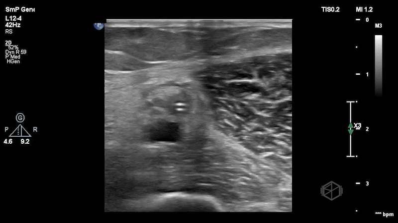

The clips here show the midline catheter in the brachial vein (paired brachial artery underneath). The catheter is surrounded by thick echogenic material - a clot, and is non-compressible. In this case, POCUS usage accurately was able to see the issue with the line.
Diagnosis: Catheter-associated deep venous thrombosis of the brachial vein
Learning points:
Deep veins are those that have a paired artery. When performing DVT scans of the upper extremity the deep veins are the: internal jugular, subclavian, axillary, brachial, radial, and ulnar veins. The basilic and cephalic veins are not deep veins, nor are any other veins that have no paired arteries.
Midlines are effective for short-term (~21 days) vascular access with a relatively low risk of complications such as lower risk of blood stream infections (35948122, 34842905).
When performing midlines, it is preferable not to use deep veins unless there is no other option. The basilic vein is the most preferred vein. The basilic and brachial are preferred over the cephalic vein as their higher flow ensures lower and the cephalic vein narrows proximally.
In one systematic review, midline catheters were associated with a higher risk of DVTs than PICCs. The authors suggest that the increased risk may be due to factors such as catheter-to-vein ratio, dwell time, and insertion technique (33991462).
Larger diameter catheters are associated with a greater risk of DVT. When placing midlines, the smallest diameter catheter necessary should be preferentially selected (37331120).
With a malfunctioning midline, POCUS can help visualize the vessel and the catheter, and can help diagnose complications such as thrombosis or obstruction.