
July SonoProps
Welcome to July’s SonoProps. This month’s theme is: Things aren’t always what they seem…
Our first SonoProps goes to Dr. Christine Hickey!
Dr. Hickey was resuscitating a male patient of unknown age who was brought to the ED in traumatic cardiac arrest after being struck by a car. The patient had pulses briefly after EMS resuscitation, but then became bradycardic and lost pulses. The patient was resuscitated in ATLS fashion and eventually Dr. Hickey noted this echo:
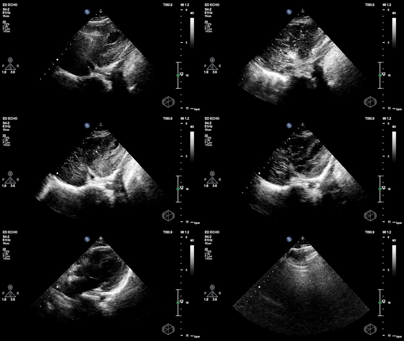
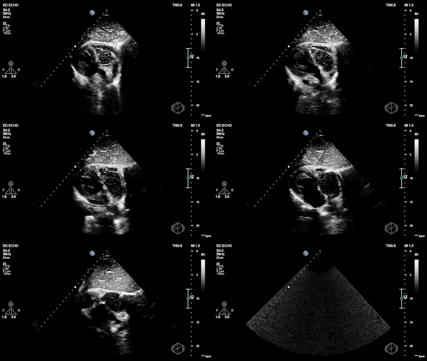
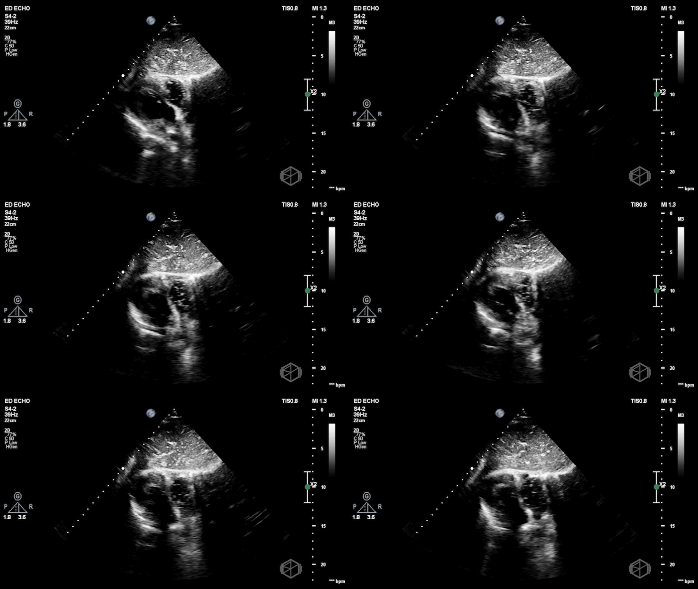
Although the patient remained pulseless, POCUS demonstrated chaotic fibrillatory myocardial activity consistent with ventricular fibrillation. The patient was immediately defibrillated, and resuscitative efforts continued. Unfortunately, despite continued aggressive management, the patient did not survive.
Diagnosis: Ventricular fibrillation
Learning points:
POCUS may uncover ventricular fibrillation that is not obvious on the monitor. This has been described as “occult VF,” where the myocardium appears to be fibrillating on ultrasound even though the surface rhythm looks like PEA or asystole.
In a 2025 multicenter study of 811 cardiac arrest patients, occult VF was seen in 5.3% of cases. Most of those patients appeared to be in PEA on the monitor, while the rest appeared to be in asystole. Key point: when the monitor and POCUS do not match, there may be an opportunity to defibrillate.
(📚 PMID: 40590825)
The monitor does not always match the mechanical activity of the heart. In a large prospective cardiac arrest study, ECG findings and myocardial activity on ultrasound were different during 28.6% of pulse checks. About one in four rhythms that appeared shockable on ultrasound looked non-shockable on the monitor and were not defibrillated. (📚 PMID: 34798178)
Do not let ultrasound prolong pulse checks. One study found that pulse checks lasted about 21 seconds when POCUS was used, compared with 13 seconds without it. A structured approach, such as the CASA protocol, can help keep pauses short. Get the probe in position and find your window during compressions, record a brief clip during the pulse check, restart CPR, and interpret the POCUS while compressions are ongoing.
(📚 PMID: 28754527, 30071262)Cardiac standstill is strongly associated with mortality, but keep in mind the overall clinical picture. A systematic review of traumatic cardiac arrest found an extremely poor prognosis when no cardiac activity was present; however, the authors determined that the available evidence was insufficient to recommend stopping resuscitation solely on the basis of POCUS. Ultrasound findings must be integrated with the mechanism, downtime, rhythm, reversible causes and the overall clinical picture. (📚 PMID: 34437998)
The next SonoProps goes to Dr. Eytan Mendelow.
Dr. Mendelow performed this focused assessment for free fluid on an approximately 40-year-old female who presented to the ED with right lower quadrant abdominal pain. The patient had a history of a prior left sided ectopic pregnancy status post left salpingectomy as well as other two other ectopics treated with methotrexate. The pregnancy status was unknown at the time of the POCUS.
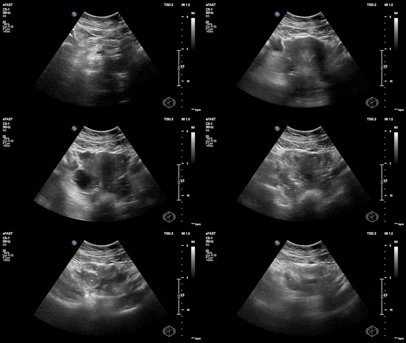
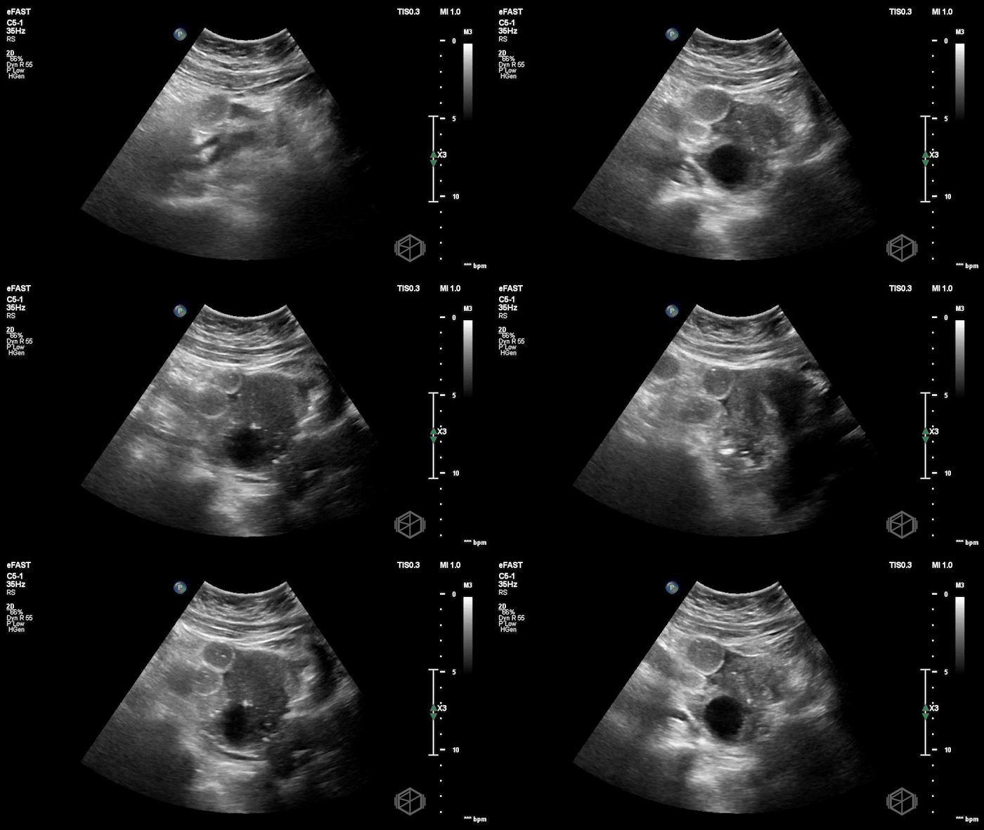
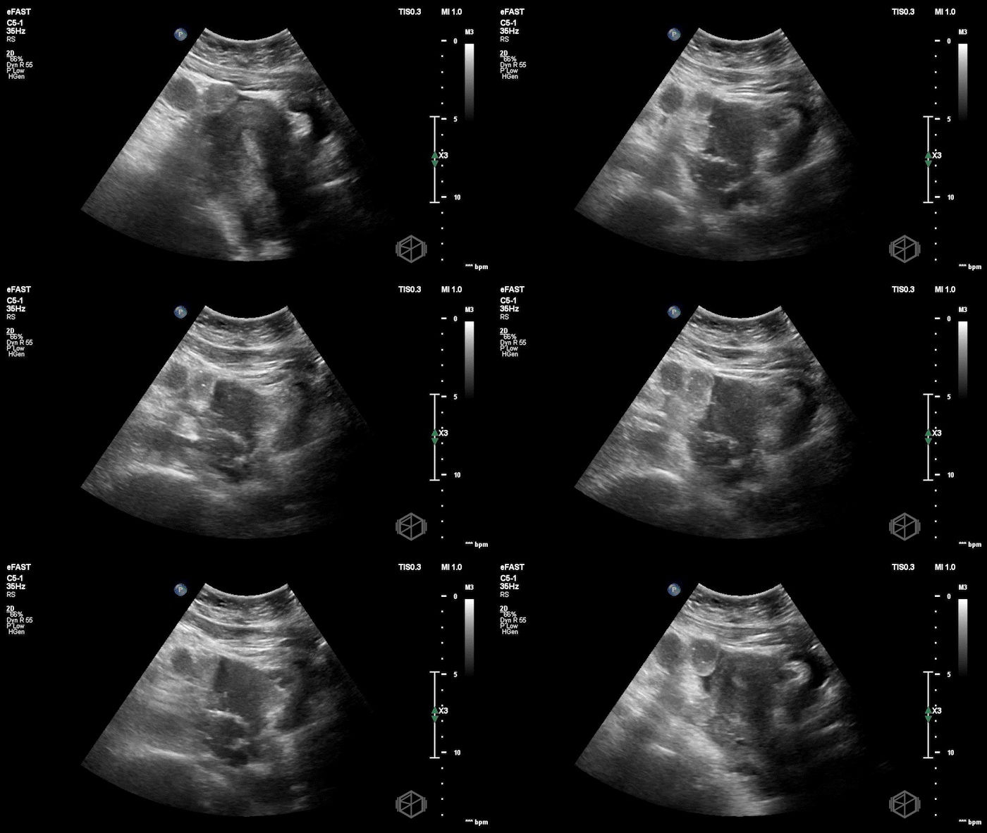
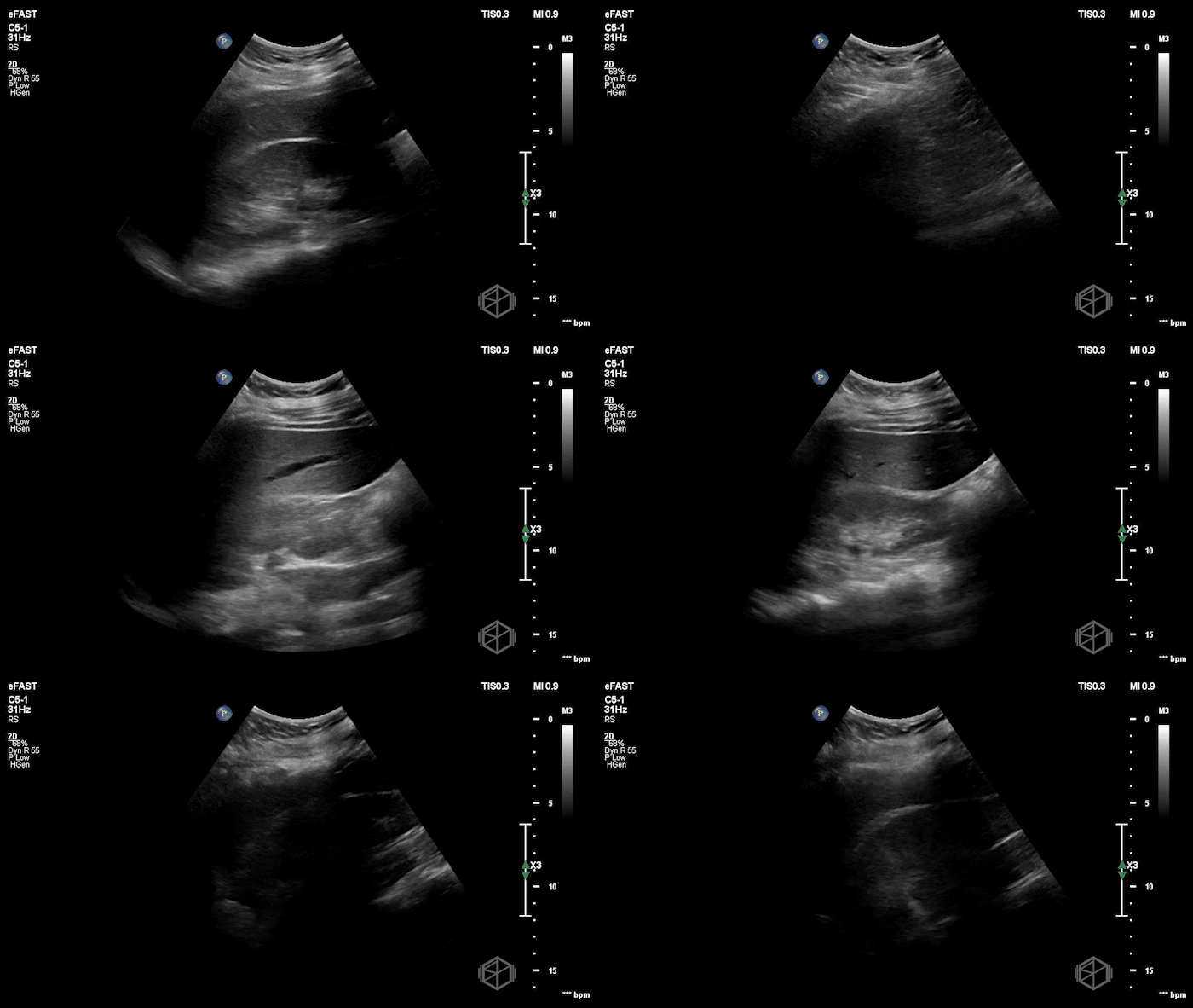
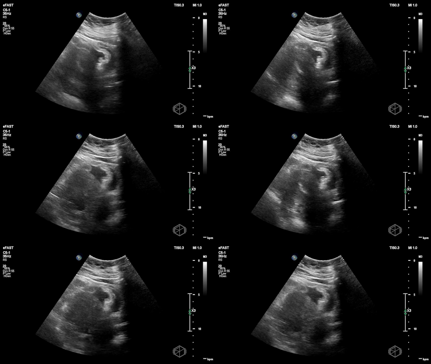
The POCUS shows free fluid behind the uterus, a right sided adnexal mass, and an empty uterus. The right upper quadrant was negative for free fluid. The patient terminated the exam due to pain. Her beta-hCG later returned at 5,900 mIU/mL.
The patient later had radiology performed transabdominal and transvaginal ultrasounds. The study was read as showing no intrauterine or ectopic pregnancy, a right hemorrhagic ovarian cyst, and no free fluid.
Given the history of prior ectopic pregnancies and the patient’s continued abdominal pain, OB/GYN took the patient to the operating room and the patient had a right-sided unruptured ectopic pregnancy with approximately 30 mL of hemoperitoneum in the posterior cul-de-sac.
Diagnosis: Right-sided ectopic pregnancy with hemoperitoneum
Learning Points:
POCUS may catch a finding early that is harder to appreciate later. The bedside exam was performed while the patient was actively having pain and was focused on the right adnexa and posterior cul-de-sac. Small amounts of free fluid and subtle adnexal abnormalities are not seen consistently on every examination. Ultrasound findings may differ between examinations because of patient positioning, the views obtained, operator technique, and the subtlety of the findings. A small amount of pelvic free fluid seen on an earlier bedside exam may not be identified on a later study. Examiner experience also affects the detection of findings. (📚 PMIDs: 25766776, 7782186)
POCUS can shorten the time to diagnosis and treatment. In a small retrospective study of patients with ruptured ectopic pregnancies, the mean time from ED arrival to ultrasound interpretation was 15 minutes with POCUS and 138 minutes with radiology ultrasound. The mean time to OB consultation was 35 versus 150 minutes, and the mean time from ED arrival to the operating room was 160 versus 381 minutes.
(📚 PMID: 34538680)Review the images when the report does not match the clinical picture. A recent case series described two patients whose POCUS exams showed adnexal masses and pelvic free fluid, while the radiology interpretations did not confirm ectopic pregnancy. Continued concern and review of the images led to operative management, where both patients were found to have ruptured ectopic pregnancies. Persistent pain, a high-risk history, and concerning bedside findings should prompt direct discussion with OB/GYN. (📚 PMID: 40620836)
An ectopic pregnancy can resemble a corpus luteum or hemorrhagic ovarian cyst. The adnexa can be difficult to interpret when a definite gestational sac, yolk sac, or embryo is not visible. Comparing the echogenicity of the mass with the ovary and confirming that the mass moves separately from the ovary may help, but the ultrasound still has to be interpreted alongside the pregnancy location, symptoms, beta-hCG, and clinical history. (📚 PMID: 11149525, 14756354)
A negative radiology ultrasound does not exclude ectopic pregnancy. Transvaginal ultrasound can miss an ectopic pregnancy when the findings are small or nonspecific. A symptomatic patient with a positive beta-hCG and no confirmed intrauterine or ectopic pregnancy still has a pregnancy of unknown location. In this case, the history of recurrent ectopic pregnancy, ongoing pain, right adnexal abnormality, and pelvic free fluid remained concerning despite the radiology ultrasound interpretation. (📚 PMID: 31618321)
