
June SonoProps
Welcome to the end of spring!
Our first SonoProps goes to Christine Cirillo, NP - our first NP to get a SonoProps recognition. Christine had a 30-year-old male presenting to the ED with cough, fever, shortness of breath and left sided chest pain. He was treated for a pneumonia outpatient with Azithromycin, then Augmentin, but despite the antibiotics his symptoms worsened.
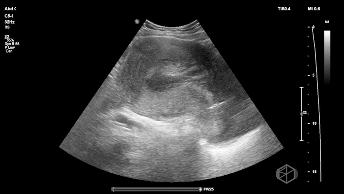
Christine obtained a POCUS immediately that demonstrated the following:
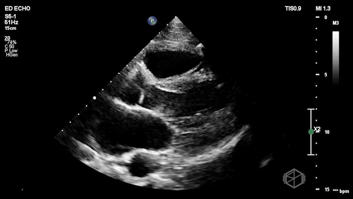
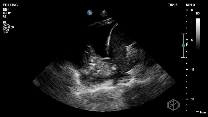
The POCUS shows an anechoic collection below the heart next to the descending aorta indicating a pleural effusion rather than a pericardial effusion. This is further confirmed by looking at the lung base where there is an obvious spine sign. There is also a consolidated lung present.
Diagnosis: Left parapneumonic effusion with complicated pneumonia
Learning Points:
Worsening dyspnea, pleuritic chest pain, persistent fever in patients with pneumonia, especially when treated with antibiotics, should prompt evaluation for a parapneumonic effusion or empyema. Parapneumonic effusions are also common in hospitalized patients with pneumonia. POCUS is more sensitive than physical exam and chest radiography for identifying pleural effusions and can help guide management at the bedside. (📚 PMID: 16493154, 26218493, 34487258, 20676576)
On lung ultrasound, pleural fluid can be described as anechoic, complex non-septated, complex septated, or homogeneously echogenic. Simple effusions are anechoic. Simple effusions can be transudative or exudative. Complex effusions can have septations, loculations, fibrin stranding, or echogenic debris. Complex effusions are exudative, indicating a complicated process such as a parapneumonic effusion, empyema, or malignancy.
Antibiotics alone may not be enough for parapneumonic effusions.
Pleural fluid pH is one of the most useful markers: lower pH, especially pH ≤ 7.2, supports complicated parapneumonic effusion or pleural infection and should prompt consideration of drainage when there is a safe accessible pocket.
Other concerning findings include frank pus, positive Gram stain or culture, low glucose, elevated LDH, large effusion, or septations on ultrasound. (📚 PMID: 7767510, 37553157)
POCUS expedited care for this patient. It rapidly identified the pleural effusion and helped shift the evaluation from treatment-failure and persistent pneumonia to complicated pneumonia with parapneumonic effusion. Early recognition helped guide next steps, including escalation of care, admission, and consideration of drainage. (📚 PMID: 26218493, 37553157)
If thoracentesis or drainage is needed, ultrasound guidance matters.
A systematic review and meta-analysis found that ultrasound guidance was associated with a lower risk of pneumothorax after thoracentesis.
(📚 PMID: 20177035)
A large database study also found ultrasound guidance was associated with fewer thoracentesis-related pneumothoraces and lower cost of care. (📚 PMID: 23381318)
The next SonoProps goes to Dr. Yanal Maher.
He had an approximately 30-year-old female presenting to the ED with vaginal bleeding and contractions. She was reportedly approximately 19 weeks pregnant. The patient unfortunately passed the products of conception including the placenta while in the ED.
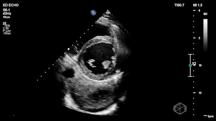
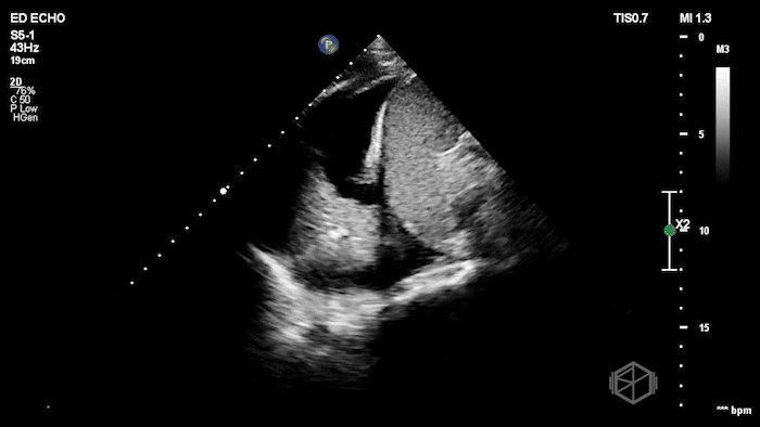
POCUS revealed the following findings afterward:
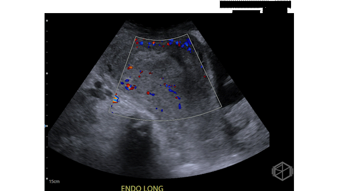
The POCUS shows a large amount of heterogeneous material within the endometrial canal consistent with retained products of conception (RPOC).
Diagnosis: RPOC
Learning Points:
In a patient with pregnancy loss, it is important to look for RPOC. Ongoing bleeding, pain, hypotension, or fever should raise concern. POCUS can help rapidly identify persistent intrauterine material and escalate care early.
(📚 PMID: 26966600, 39826241)RPOC often appears as a heterogeneous or echogenic mass within the endometrial cavity.
(📚 PMID: 16123177, 33459841)Color Doppler can be helpful:
Vascularity within the endometrial material increases concern for retained products
Avascular heterogeneous material may represent clot.
Absence of Doppler flow does not exclude RPOC.
Endometrial thickness can support the diagnosis, but it should not be used in isolation. An endometrial mass is the most sensitive (79%) and specific (89%) sonographic feature for RPOC. If no endometrial mass or fluid is seen and the endometrial thickness is less than 10 mm, RPOC is unlikely.
(📚 PMID: 16123177)POCUS is useful for ruling in concern for RPOC, but it is not perfect for ruling it out. A recent ED study found that POCUS had high specificity (93.8%) and moderate sensitivity (79.0%) for diagnosing RPOC.
(📚 PMID: 39826241)In second-trimester pregnancy loss, retained placenta or retained products can be a major driver of hemorrhage. After delivery or passage of products, POCUS can help guide the next steps, including uterotonics, transfusion planning, expediting OB consultation, and possible uterine evacuation.
(📚 PMID: 33459841, 31006292)