
July SonoProps
SonoProps #1 goes to... Dr. Mauren Aiad!
A 38-year-old female presented to the emergency department with abdominal pain with a positive pregnancy test.
Dr. Aiad promptly took the ultrasound over to the patient and did a FAST and saved images that showed this:
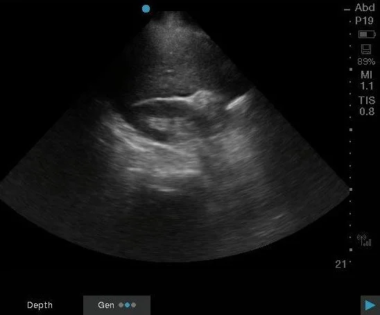
This still image shows a sliver of free fluid within the hepatorenal space and potentially some free fluid near the liver edge. Dr. Aiad had the answer quickly: concern for a ruptured ectopic pregnancy.
Within 15 minutes Ob/Gyn was at the bedside. Later, the radiology US showed: "No intrauterine gestational sac identified. Right adnexa is suboptimally evaluated. There is a moderate amount of pelvic free fluid. This is nonspecific and, although this could be secondary to a ruptured hemorrhagic ovarian cyst, ruptured ectopic pregnancy cannot be excluded." HCG was 1378.
The patient was taken to the OR later and noted to have "300 mL of hemoperitoneum in the pelvis upon entry into the abdomen. Ruptured ectopic pregnancy noted in the ampulla of the right fallopian tube."
Excellent job Dr. Aiad.
Learning Points:
Any female with abdominal pain/vaginal bleeding in the setting of a positive pregnancy test will benefit from a quick FAST and a transabdominal pelvic US (and remember to save clips, no ghost scanning!). Sensitivity increases if there is hypotension present.
There is no specific HCG level for an ectopic pregnancy to be present. Ruptured ectopic pregnancies can present with extremely low HCGs (even as low as less than 10 mIU/mL)
An empty uterus with free fluid up to the RUQ in the setting of a positive pregnancy test is concerning for a ruptured ectopic pregnancy until proven otherwise.
The most sensitive site for detection of free fluid in the RUQ is not actually Morrison's pouch but the caudal liver edge.
SonoProps #2 goes to... Dr. Christopher Scavelli!
A 72 year old female presented to the ED with chest pain. Dr. Scavelli took the ultrasound and performed a cardiac POCUS and saved clips/images which showed this:
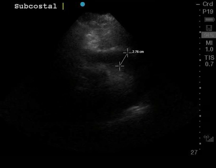
The patient had just had an echo very recently that had NO pericardial effusion. The clip demonstrates a large circumferential pericardial effusion in a parasternal short view. The heart looks like its floating in fluid.
Dr. Scavelli noted that the patient was hypotensive and given the POCUS had a pericardial effusion which equated to possible tamponade, he immediately called CT surgery who agreed with his assessment. The patient went to the OR and had 550cc of bloody pericardial fluid drained.
Great job Dr. Scavelli.
Learning Points:
Cardiac tamponade is a clinical diagnosis, but ultrasound findings precede clinical tamponade.
Acuity matters more than size.
Hypotensive patients will benefit from a cardiac POCUS (which includes the IVC) at the least, but depending on the patient other areas to consider include the lungs/thorax, a FAST, and aorta. You do not have to spend time doing all the scans, use your clinical judgement.
Features of cardiac tamponade on ultrasound include right atrial systolic collapse, right ventricular diastolic collapse, a plethoric IVC, and a mitral inflow variation >25%.
A new pericardial effusion in a patient with hypotension is concerning for tamponade.