November SonoProps
November is here and so are the start of the holidays.
This month’s first SonoProp goes to Dr. Vivek Sharma for a holiday story gone wrong.
Dr. Sharma responded to a level 2 trauma for a 75-year-old male who fell down a ladder about 5 steps tall while hanging decorations onto his left side. The patient was able to get back up and stand. He had no external wounds or injuries except for left anterior rib tenderness.
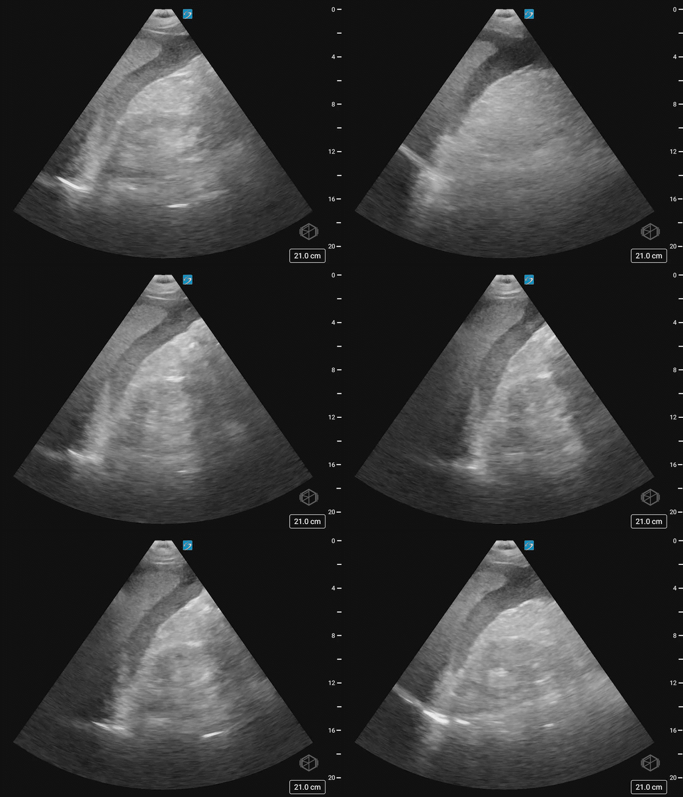
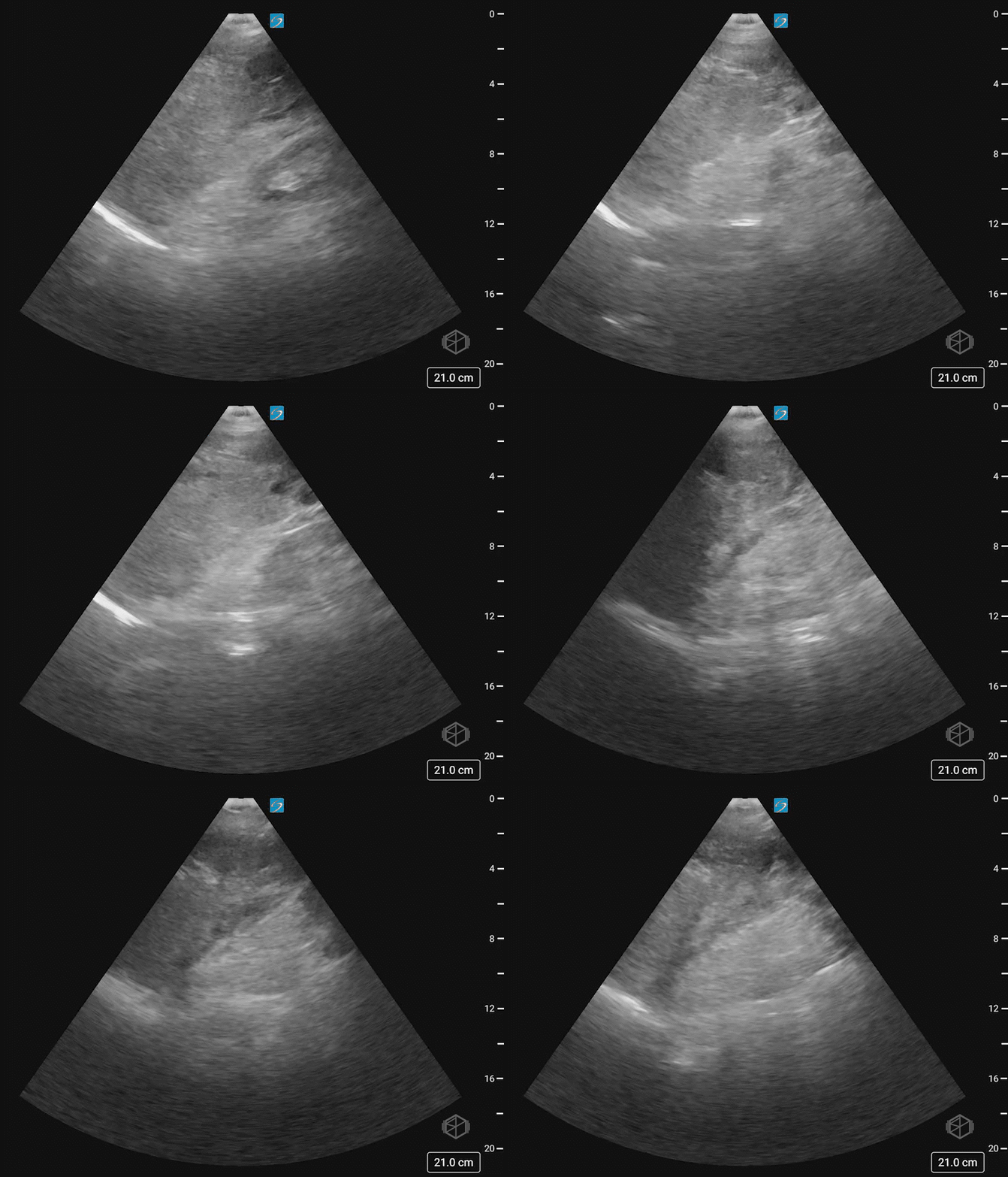
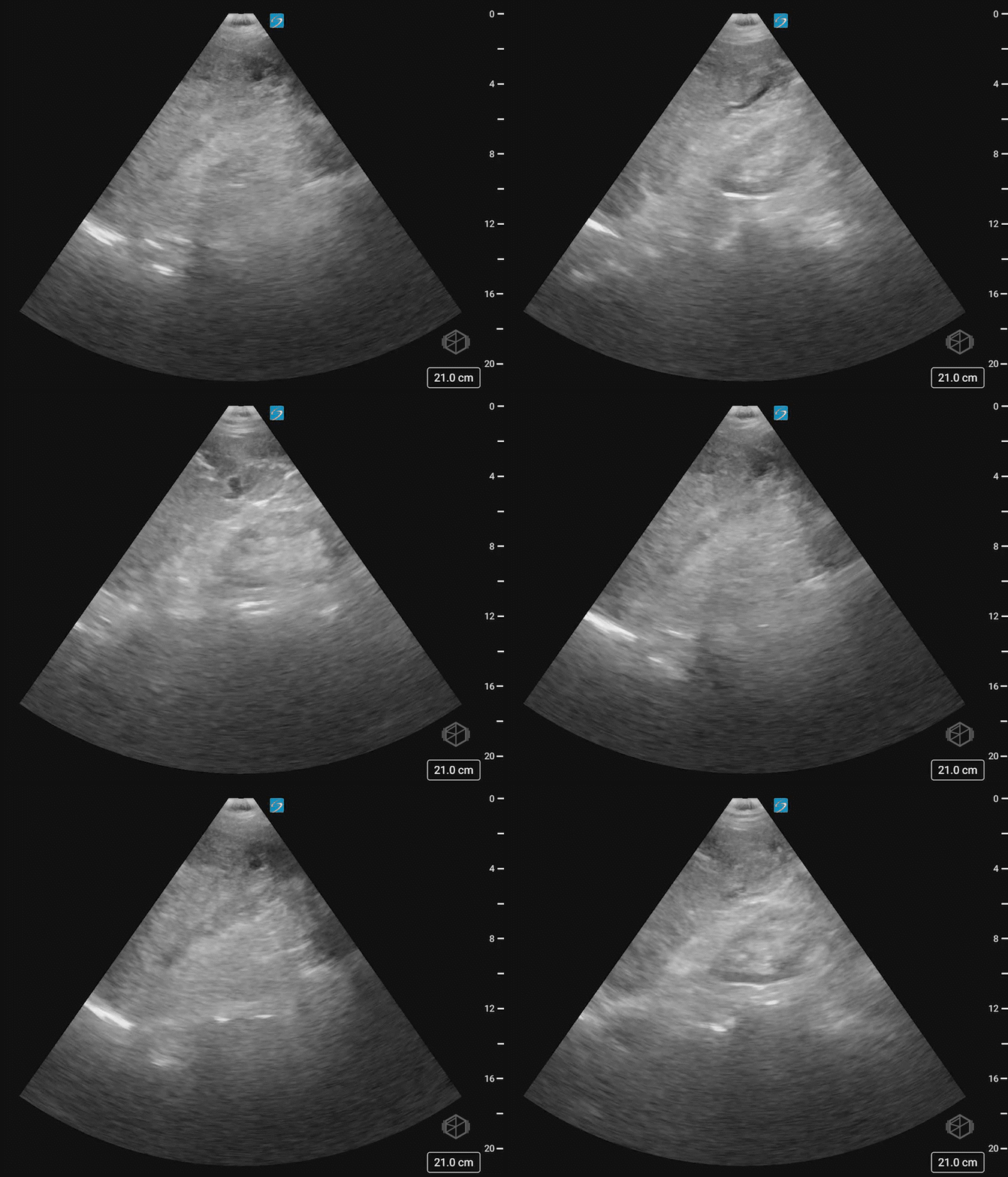
This was the patient’s FAST examination:
This FAST examination is clearly positive, with large amounts of complex fluid near the liver tip in the first clip. In the 2nd and 3rd clip, note the absence of the normal contour of the spleen. There is also free fluid at the tip of the spleen.
The patient had a CT that demonstrated AAST spleen grade grade 5 splenic injury based on active bleeding with hemoperitoneum and splenic laceration and left fourth through eighth rib fractures.
Diagnosis: Grade 5 splenic laceration with hemoperitoneum and active extravasation
Learning points:
Swirling internal echoes in this scan likely indicate active hemorrhage.
When evaluating the left upper quadrant, remember the spleno-diaphragmatic space (28210364) and the inferior paracolic gutter (26206829) have been found to be the most sensitive areas for free fluid.
Isolated left upper quadrant free fluid is found mostly in the subdiaphragmatic space due to the phrenicocolic ligaments. The phrenicocolic ligament limits fluid movement from the left paracolic gutter to the left flank, which lets fluid migrate across the midline into the right flank. With large fluid volumes there may be accumulation between the spleen and kidney. Remember to scan all the way from above the diaphragm to below the kidney.
Normally the spleen on ultrasound is smooth and homogeneous. Evaluating the heterogeneity of the spleen can indicate splenic injury (36601219, 23175018).
In one study, sensitivity for detection of splenic injuries was 86% for grade III or higher injuries. Ultrasound is most sensitive for the detection of grade III or higher BSI based on the presence of haemoperitoneum (11223039).
The next SonoProp for this month goes to our current resident rotator Dr. Eytan Mendelow.
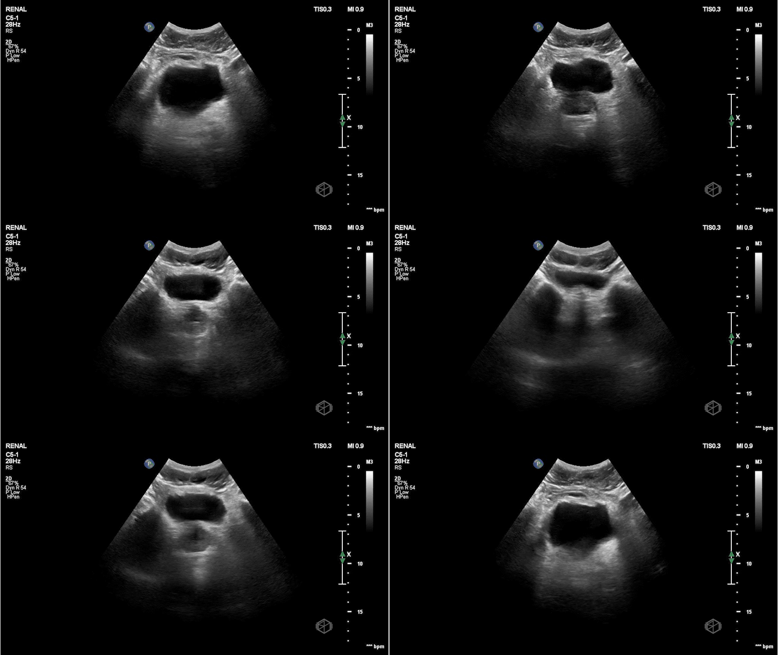
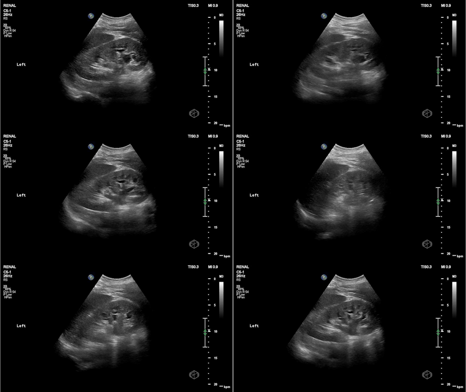
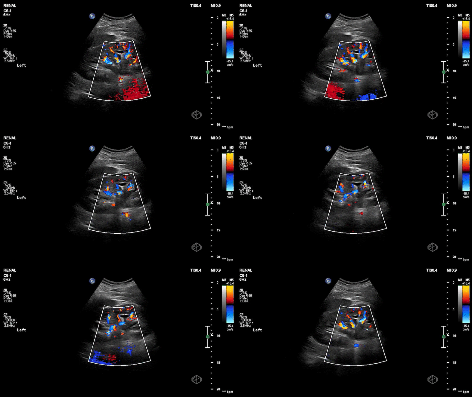
As you know, during the holidays, delivery drivers work extremely hard to get our packages to us as quickly as possible. Dr. Mendelow scanned a middle-aged delivery man with left flank pain that came on suddenly while he was out delivering holiday packages. The right kidney was normal. He obtained the following images:
This patient has mild-to-moderate left hydronephrosis with an approximately 5mm ureteropelvic junction (UPJ) stone. Note the hyperechoic stone with shadowing posterior to it just outside the kidney in image 4 and in clip 2. The patient later had a CT that demonstrated the same but the stone had moved just slightly lower into the proximal ureter.
Diagnosis: Left mild-to-moderate hydronephrosis with a UPJ stone
Learning points:
Them most common sites to visualize obstruction for kidney stones are the ureteropelvic junction (UPJ) and the ureterovesicular junction (UPJ).
Grading of hydronephrosis goes from mild to severe. The actual interpretations are varied and often times it’s perfectly fine to say mild-to-moderate or moderate-to-severe as it can be difficult to delineate (32984198).
Mild hydronephrosis involves only the renal pelvis and some calyces but the pelvico-calyceal architecture is maintained.
Moderate hydronephrosis begins to involve the medullary pyramids and there is an outpouching of the calyces, sometimes called the “cauliflower” appearance.
Severe hydronephrosis — the renal pelvis and calyces are ballooned and the there is cortical thinning.
It is important to when hydronephrosis is noted to look at the ureteropelvic junction, as subtle stones may be missed. Especially if the bladder scan does not demonstrate any hyperechoic stones or “twinkling” artifact.

