October SonoProps
October brings many things, Fall, Halloween, many other cultural holidays… but it also brings us another edition of SonoProps.
Our first SonoProps goes to Dr. Rachel Ariz!
Dr. Ariz was doing an echo on a 61-year-old male with a history of hypertension and CKD who presented to the ED with fever, shortness of breath, and vague abdominal pain.
The patient’s scan demonstrated the following:
This patient has a large circumferential pericardial effusion. There is early diastolic collapse of the right ventricle, and early right atrial systolic buckling. The RV and RA are not filling entirely when they’re supposed to due to external pressure from the pericardial effusion. The patient was hypertensive and tachycardic, so not clinically in tamponade.
Dr. Ariz notified the attending who called cardiothoracic surgery (TCV), cardiology, and the ICU. On ICU’s echo patient had a mitral inflow variation of 42% (another sign of tamponade).
TCV noted the patient was "stable" and recommended watchful waiting. However, the ICU attending opted to perform a pericardiocentesis and placed a pericardial drain. They explained their decision with a clear analogy:
“When you smell a gas leak in your house, you don’t wait for flames to appear before turning off the gas and calling the gas company. You act immediately.”
Similarly, waiting for a patient to develop critical tamponade before intervening may not be the safest approach. It's better to act while the patient is semi-stable, especially when multiple sonographic signs of tamponade are present.
Additionally, the patient had acute renal failure and a possible infectious process, making it crucial to distinguish between uremic and infectious pericardial effusion.
Diagnosis: Large pericardial effusion with multiple signs of tamponade
Learning points:
As with earlier SonoProps - there are 4 key signs of the EM physician to know for sonographic tamponade: RA systolic collapse, RV diastolic collapse, a plethoric IVC, and mitral inflow variation >25%.
Sonographic tamponade signs precede clinical signs. Remember, just because a patient is stable with signs of early tamponade on echo, does not mean they’ll stay stable.
Pericardial drainage for 24-72 hours is generally sufficient to prevent recurrence of tamponade in a majority of cases (21907951, 24399485).
There are many causes of pericardial effusions:
Idiopathic pericarditis
Infection (bacterial or tubercular pericarditis)
Trauma
Uremia
Hypothyroidism (myxedema)
Autoimmune — Dressler’s, Lupus
Post-radiation
The next SonoProp goes to Dr. Frances Rusnack.
Dr. Rusnack had a 62-year-old male patient with no significant past medical history with intermittent right hand numbness that started after using a wrench 4 weeks ago. The patient was seen at an urgent care and diagnosed with tendinitis. On the day of presentation noted that this hand was cold and paler compared to his left hand.
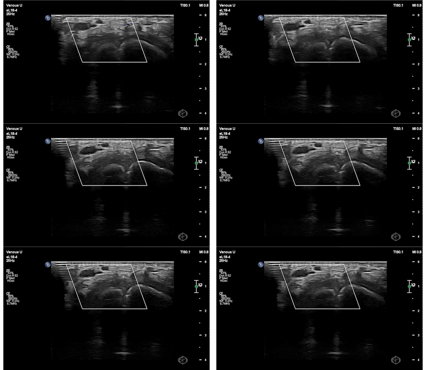
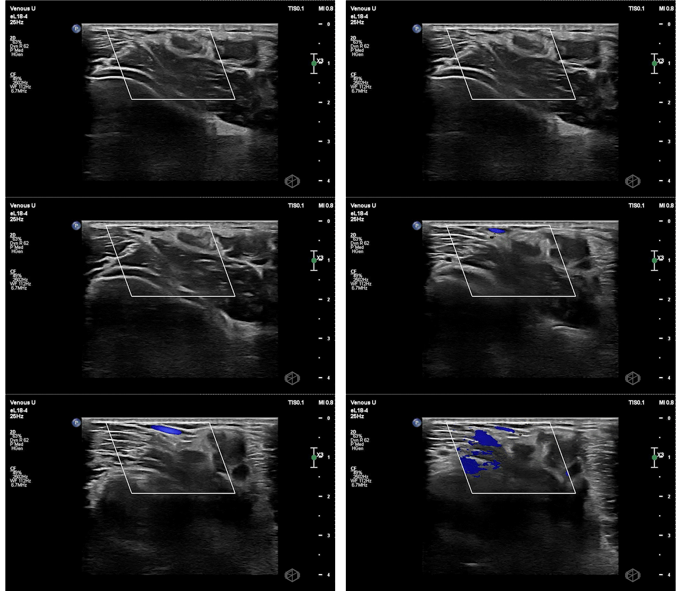
Dr. Rusnack performed a POCUS of the right arm that demonstrated the following:
In clip 1, the patient has no doppler flow noted right over the radial artery. On clip 2, the patient has venous flow but no notable pulsatile flow directly over the artery. The artery also appears mildly enlarged with a slightly edematous appearing wall. Vascular surgery was immediately consulted, heparin was started, and CTA demonstrated a proximal to mid brachial artery occlusion. The patient was taken to the OR by vascular surgery for an embolectomy and bypass. A simple scan, that took less than a few minutes to do.
Diagnosis: Acute upper extremity non-traumatic arterial occlusion
Learning points:
There are few case reports of unprovoked brachial artery occlusion (37152341, 36427028, 36427028).
Nontraumatic upper extremity acute limb ischemia (ALI) is a limb and potentially life-threatening pathology with an estimated incidence of about of 1.2 to 3.5 per 100,000 per year, or approximately 1/5th that of acute leg ischemia (9782011).
A patient with acute limb ischemia typically presents the 6P’s: paresthesia, pain, pallor, pulselessness, poikilothermia, and progression to paralysis.
POCUS is a great initial test for acute arterial occlusion, however if signs of occlusion are not identified and suspicion is high enough, additional testing should be obtained. It is specific but not as sensitive as CTA or MRA (17548364).


