
September SonoProps
September brings back to school and so this month’s SonoProps theme is pediatrics!
Our first SonoProp goes to Dr. May Ali. Dr. Ali was doing a scan on an 11-year-old male with RLQ pain. The patient had minimal diffuse abdominal tenderness and right lower quadrant tenderness. The following are clips from the scan:
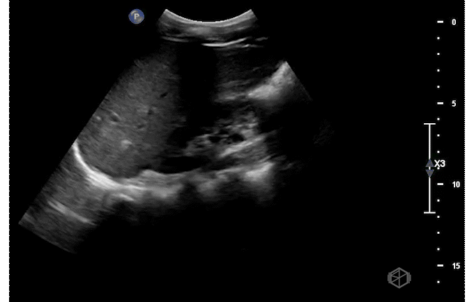
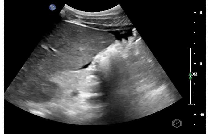
The scans show small-to-moderate free fluid in the abdomen. The appendix is not visualized. The patient had a radiology ultrasound that demonstrated a partially visualized normal appearing appendix with no ancillary findings to suggest appendicitis. Small volume free fluid in the right lower quadrant with small volume perihepatic free fluid. Labs were normal with a normal ESR and CRP. The patient was evaluated by surgery and discharged home with close follow up with his primary care doctor.
Diagnosis — Pediatric abdominal pain with free fluid, likely gastroenteritis
Learning points:
Although free fluid could be investigated with cross-sectional imaging, given this patient had normal labs, ESR, and CRP, a more conservative approach is reasonable.
In one study of 250 children with acute abdominal pain in comparison to 50 asymptomatic children, free fluid was noted in 29% of symptomatic children and 6% of asymptomatic children (8301714).
A specific diagnosis was established in 39 (54%) symptomatic children.
Ultrasonography suggested the correct diagnosis in 29 out of 39 (74%) symptomatic children in whom a specific diagnosis was established at the time of discharge from the hospital such as: appendicitis (most common), enteritis, pancreatitis, ovarian cysts, PID, adnexal torsion etc.
The discharge diagnosis in the remaining 33 (46%) children was abdominal pain or gastroenteritis of unknown origin.
In another study (23980214), pediatric patients with free fluid who did not have a specific diagnosis on imaging, the mere presence of fluid and the fluid volume were not predictive of a surgical outcome.
All patients with free fluid and no associated condition who were discharged from the ED did not return with adverse outcomes.
2 patients with equivocal sonographic findings with free fluid had appendicitis, but also had physical exams highly suggestive of appendicitis.
Free fluid in pediatric patients with acute non-traumatic abdominal pain without a specific diagnosis suggested by ultrasonography may not be a significant finding if the physical exam and labs are normal.
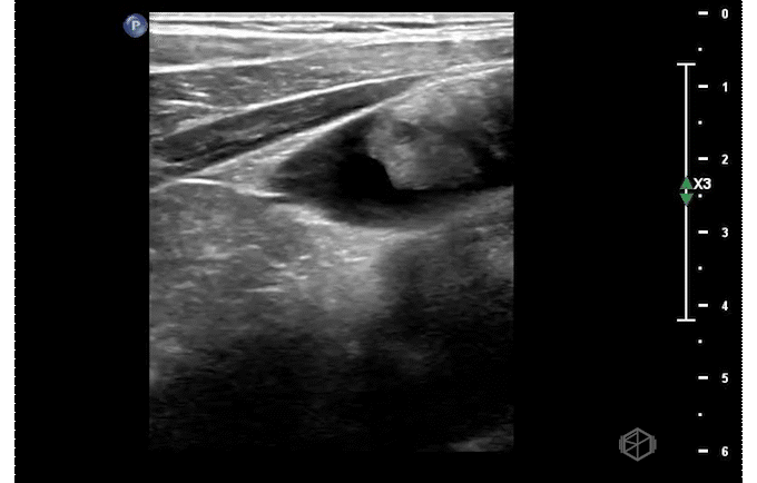
Our second SonoProp goes to Dr. Siri Tummala. Dr. Tummala scanned a 4-year-old male with LEFT elbow pain after injuring himself while going down a slide. Using the linear probe she obtained two views of the supracondylar area as seen below and compared it to the right elbow.
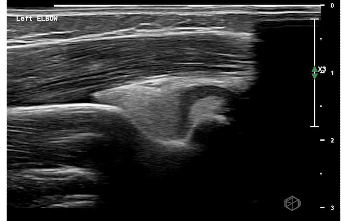
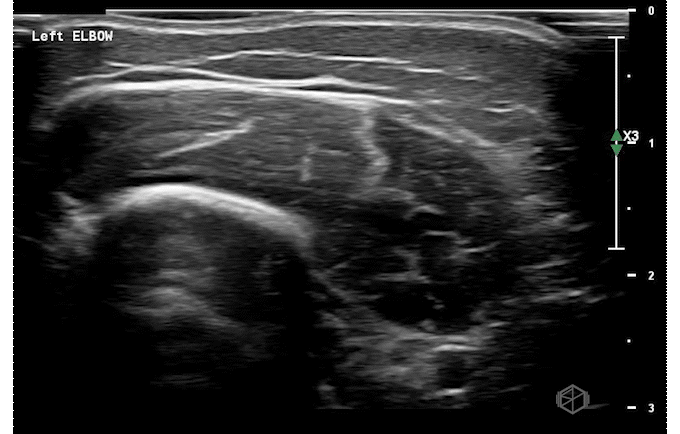

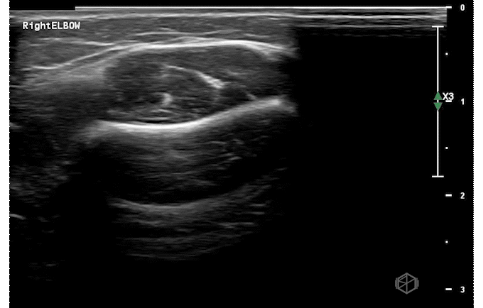


The LEFT elbow demonstrates an elevated fat pad with heterogeneous material within it representing lipohemarthrosis, while the right elbow demonstrates a normal posterior fat pad.
Diagnosis — Left elbow supracondylar fracture
Learning points:
Supracondylar fractures are the most common type of elbow fracture in pediatric patients. They can be classified using the Gartland system. Gartland Types II - IV are fairly obvious on X-ray as they are displaced. Gartland Type I fractures are non-displaced fractures, and may be missed on X-ray. A “sail sign” or a prominent posterior fat pad may provide hints that an occult fracture is present.
Ultrasound has high sensitivity (98-100%) and specificity (70-93%) for a supracondylar fractures (23142008, 26815896, 27277459). Lipohemarthrosis, a prominent dorsal fat pad are highly indicative of supracondylar fractures. Cortical disruption can be seen as well. When XR and US are both negative, the possibility of fracture can be rule out definitively (27697406).
While it does not replace X-ray, ultrasound can be used when there is a high suspicion and the X-ray is equivocal or read as negative to diagnose occult supracondylar fractures.