August SonoProps
Hello everyone,
Welcome to the August SonoProps!
Our first SonoProps this month goes to Dr. Hardeep Singh!
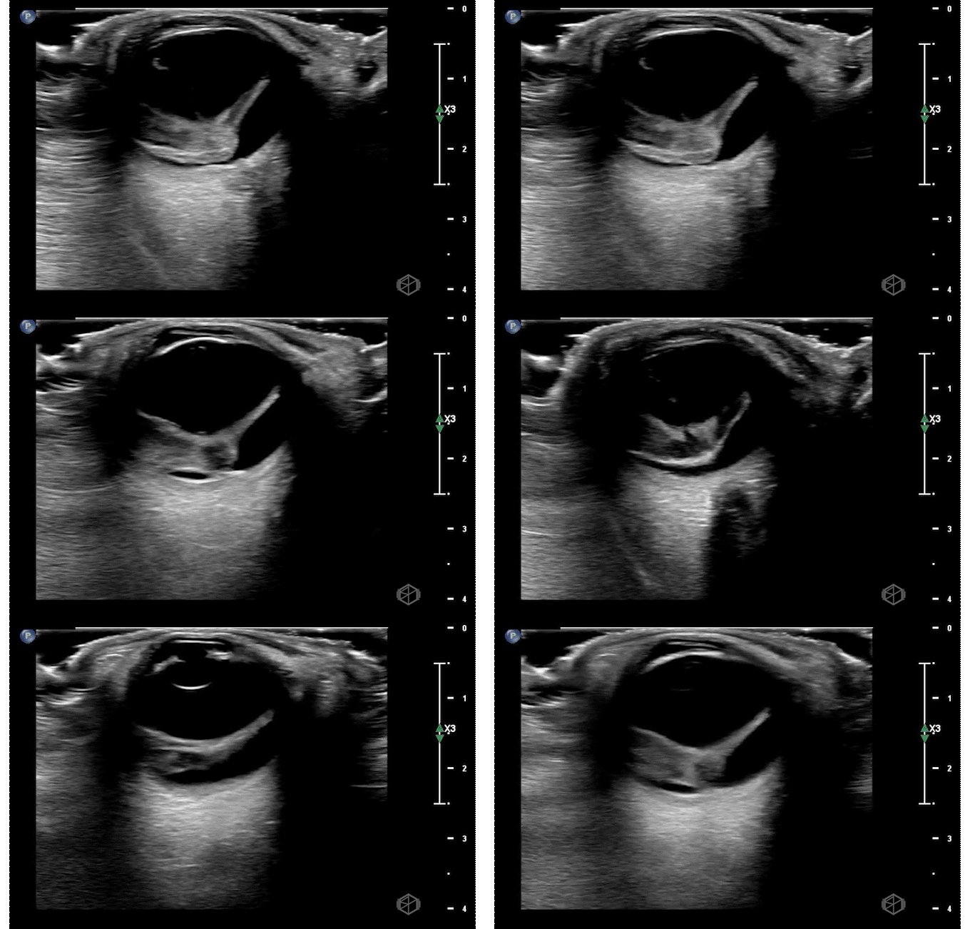
Dr. Singh had an 80-year-old female present for right eye vision loss. The patient was called a stroke alert. However, Dr. Singh was not entirely convinced it was a stroke and went one step further — he did a POCUS of the patient’s right eye and saw the below findings:
This patient has heterogeneous echogenic material within the vitreous body. There is no obvious thick membrane visible attached to the optic nerve making retinal detachment unlikely.
Diagnosis — Right eye vitreous hemorrhage
Learning points:
Vitreous hemorrhage can be due to abnormal vessels (eg, diabetes, sickle cell, retinal vein occlusion etc.), rupture of normal vessels (trauma, vitreous detachment, coagulopathy, Terson’ syndrome — SAH causing increased ICP etc.), or blood from an adjacent source.
The vitreous humor is an avascular gel-like substance that is between the lens and the retina made up of ~99% water and ~1% collagen and hyaluronic acid; vitreous hemorrhage occurs when extravasated blood enters the vitreous humor of the eye (9265701).
Appearance can depend on age of the hemorrhage — fresh hemorrhage can be wispy and mobile mildly echogenic opacities in the vitreous body. May appear more “washing machine like.” At this stage it is very important to evaluate with higher gain and perform oculo-kinetics to see the subtle hemorrhage or if a membrane present as well.
It is very important to evaluate for retinal detachment as this increases the urgency for treatment.
As the hemorrhage becomes older, it is possible to see organized echogenic opacities that layer with gravity (as seen in this example) (30977855).
The next SonoProp for this month goes to our current resident rotator Dr. May Ali.
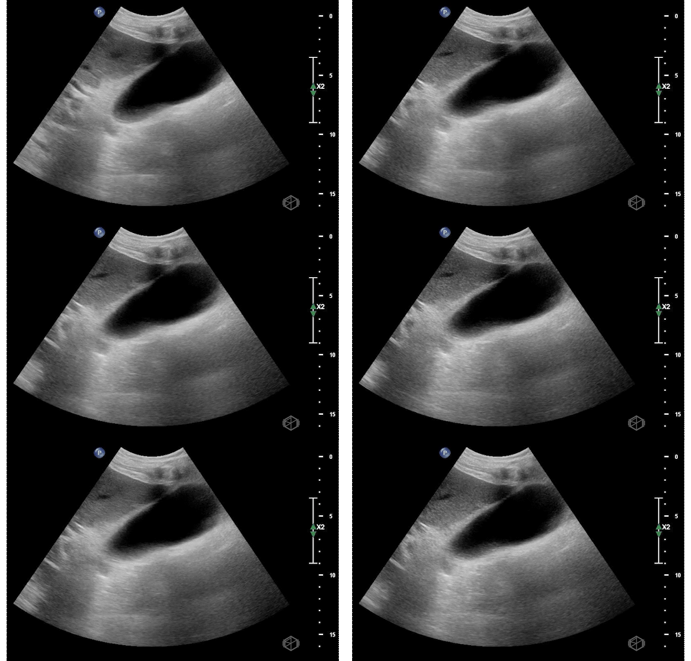
Dr. Ali was scanning an 80-year-old male patient with abdominal pain, nausea, and vomiting.
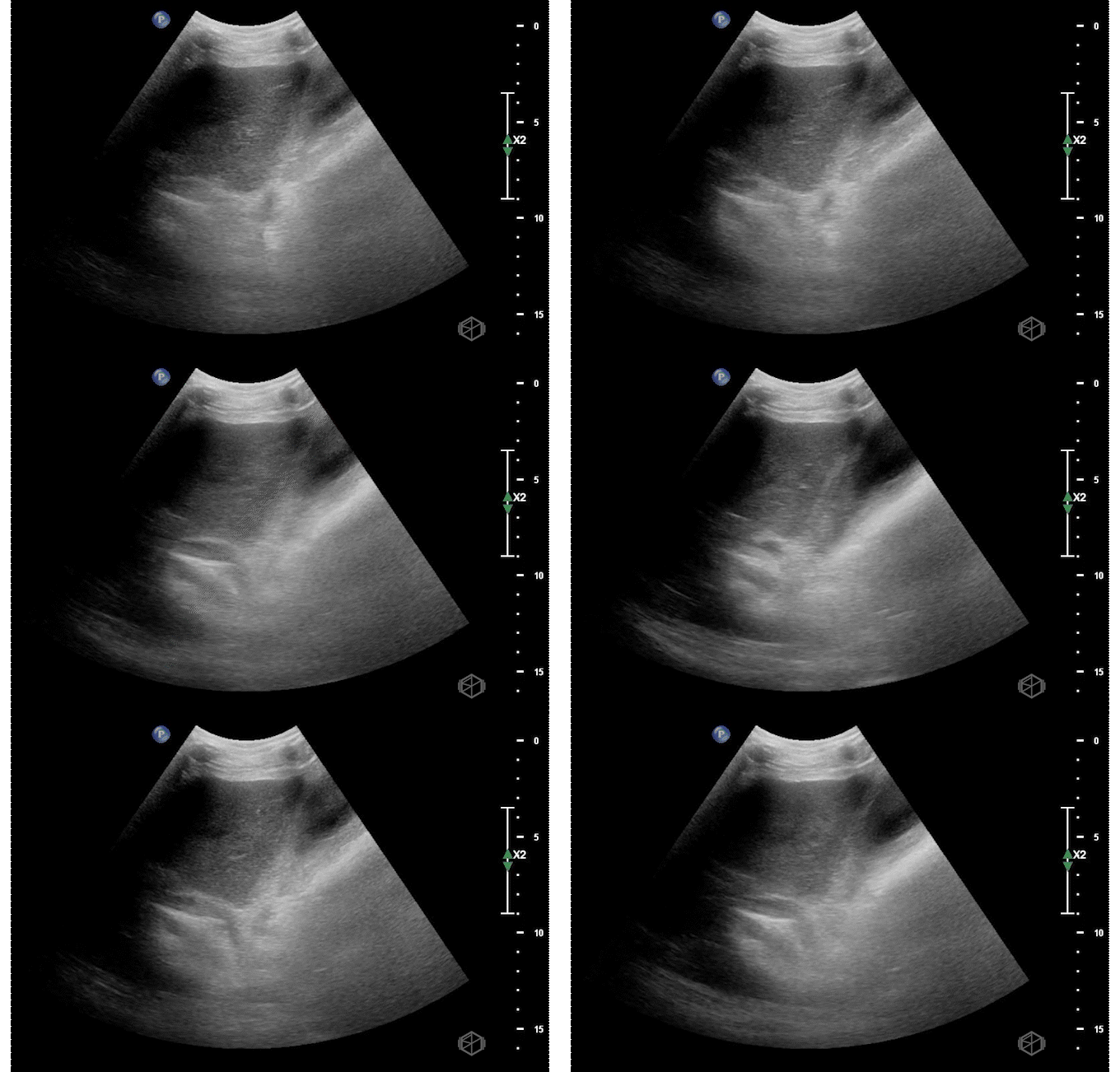
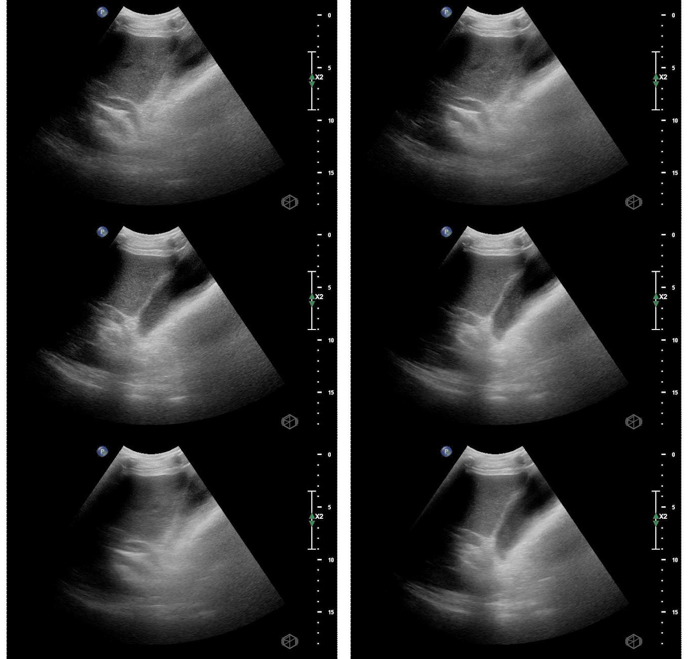
Ultrasound of the right upper quadrant demonstrated the below:
This ultrasound demonstrates a dilated common bile duct with mild gallbladder wall thickening.
The patient did have elevated liver function tests. He had an MRCP that did not identify any stones but noted some gallbladder wall thickening. Endoscopic ultrasound was negative for stones as well, but ERCP was done which noted a filling defect consistent with a stone and sludge which was removed. The patient ultimately had their gallbladder removed and pathology demonstrated acute on chronic cholecystitis.
Diagnosis — dilated common bile duct, choledocholithiasis
Learning points:
Finding the common bile duct is often difficult for novice sonographers. The common bile duct lies on top of the portal vein in long orientation making a “double barrel” type configuration as seen above. Color doppler may be used to determine the duct as there should be no flow. This is often easier to identify than the “Mickey Mouse” in short orientation.
Due to the difficulty in determining the difference between the common hepatic duct and the common bile duct, some prefer to just call this structure the common duct (504652).
If the labs are normal and there is no gallbladder wall thickening, pericholecystic fluid, or sonographic Murphy’s — there is little utility in finding the CBD (29162442).
The CBD is measured inner to inner. There are various average sizes and no standard cut off but a common rule of thumb is that a dilated common bile duct is considered >4mm, increasing by 1 every decade at 50 and after. So a 70-year-old can have up to a 7mm sized CBD (14510259).
Later studies have questioned this, however it is still considered a rule of thumb (28668951). In general, the CBD should remain under 6-7mm which is commonly accepted range for normal (11065260).


