
July SonoProps
Summer is in full swing and so is another edition of our SonoProps.
The first of this month’s SonoProps goes to Dr. Bena Mehta!
Dr. Mehta had a 74-year-old male with a history of mild COPD and active smoker who presented in with persistent worsening cough over 2 weeks with fever at home. The patient had a new requirement for supplemental oxygen.
Before even the chest x-ray Dr. Mehta did a lung ultrasound (LUS) that demonstrated the following:
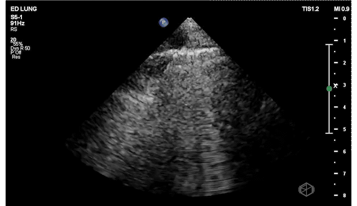
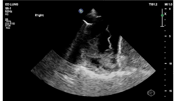
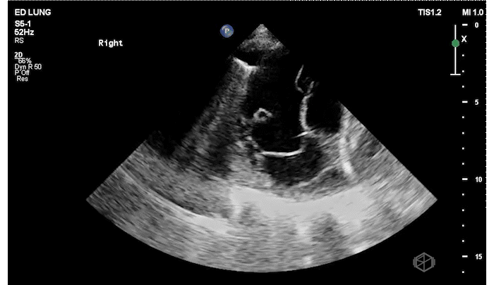
The patient has focal B-lines on the right side with diminished lung sliding. The presence of B-lines rules out pneumothorax. The right lung base has at least a moderate sized complex pleural effusion with septations.
The chest X-ray, performed later was read as: “New right lower lobe opacity, suspicious for pneumonia. Left lung is clear. Suspected trace bilateral pleural effusions. No pneumothorax. Lungs are hyperinflated, compatible with COPD.”
Diagnosis: Complex pleural effusion, empyema
CT scan performed a day later demonstrated: “complex moderately-sized verging on large right pleural effusion which is an empyema, with associated atelectasis/pneumonia of the right lower lobe,” which we already knew and the patient had a pig tail placed that put out purulent material.
Learning points:
LUS is a highly sensitive and specific test to evaluate for the presence of a complicated pleural effusion, perhaps even better than CT, and may detect parapneumonic effusions which precede empyema earlier. (27613540, 14695718)
Complex pleural effusions are the opposite of simple. Simple pleural effusions are purely anechoic and are most often transudative.
Complex effusions may have septations, fibrinous stranding, loculations, and may be homogenously or heterogeneously echogenic. They are most often exudative. (26218493)
In patients with empyema, one study evaluated patients with 4 characteristics on LUS - (I) complex non-septated effusions (plankton sign), (II) complex fixed septated effusions, (III) complex mobile septated effusions, and (IV) homogenously echogenic effusions (hematocrit sign). They found that patients with types I and IV had success with only tube thoracostomy while type II and III required further interventions.
Another study also noted that patients with septations on LUS had longer hospital stays and higher necessity for further interventions such as intrapleural fibrinolytics or surgery (11127008).
Great job Dr. Mehta, I look forward to seeing more great scans!
The next SonoProp goes to Dr. Nina Vazquez.
Dr. Vazquez had a 78-year-old female with a history of a recent pacemaker placement present to the ED for shortness of breath and left arm pain. The patient had a new pericardial effusion (which was the real reason for her shortness of breath) as well as the below finding in her LEFT upper extremity.
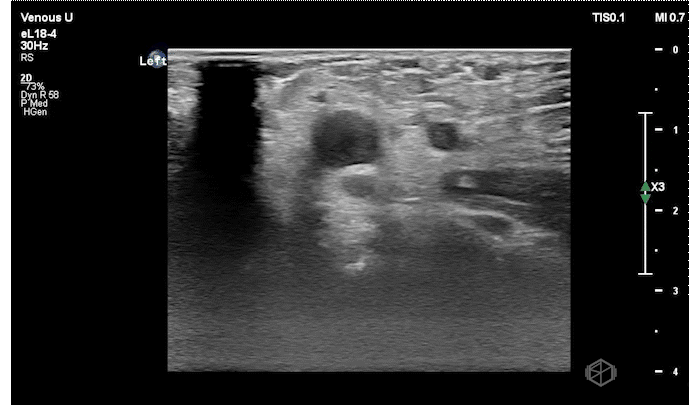


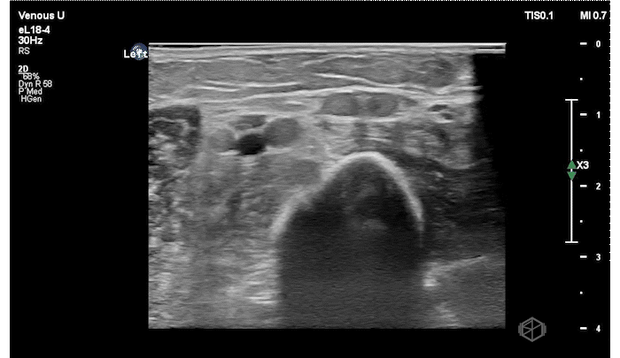
The patient has an echogenic thrombus visible in the LEFT distal subclavian vein to the branch point at the anterior cubital fossa.
Diagnosis: Left upper extremity DVT
Learning points:
Upper extremity DVTs are rare, but can have the same complications such as pulmonary embolism, post-thrombotic syndrome, and septic thrombophlebitis.
At the novice level POCUS can be used to rule in UE DVTs to expedite care, however if suspicion is high and further testing should be obtained.
Upper extremity DVT POCUS evaluates the following areas — internal jugular → brachiocephalic (BCV) & subclavian (SCV) (not easily compressible, requires pulsed-wave doppler evaluation) → axillary vein → brachial veins → radial and ulnar veins. The basilic and cephalic veins are superficial veins. (15914687, 34736403)
As with lower extremity DVT POCUS the signs of a DVT are: direct visualization of thrombus, lack of compressibility, lack of doppler signal within the vein, lack of augmentation (lower value, especially single point), lack of respiratory variation (spectral doppler, useful for SCV, BCV).