
Welcome to our November SonoProps! Our theme this month is pediatrics!
The first SonoProps this month goes to Drs. Steven LaFever and Eytan Mendelow! They were working with our fellow Dr. Brian Tang and US faculty Dr. Michael Zimmerman.
A teenage male with no significant past medical history presented as a transfer from an outside emergency department for left upper extremity pain and swelling. His symptoms began approximately two days earlier with a sensation of tightness and squeezing in his left upper arm, accompanied by swelling, paresthesias, and purple discoloration.
Interestingly, the patient was an avid violin player. Although right-handed, he held the violin with his left arm while using his right hand to control the bow.
POCUS of the left upper extremity was performed, demonstrating the following:
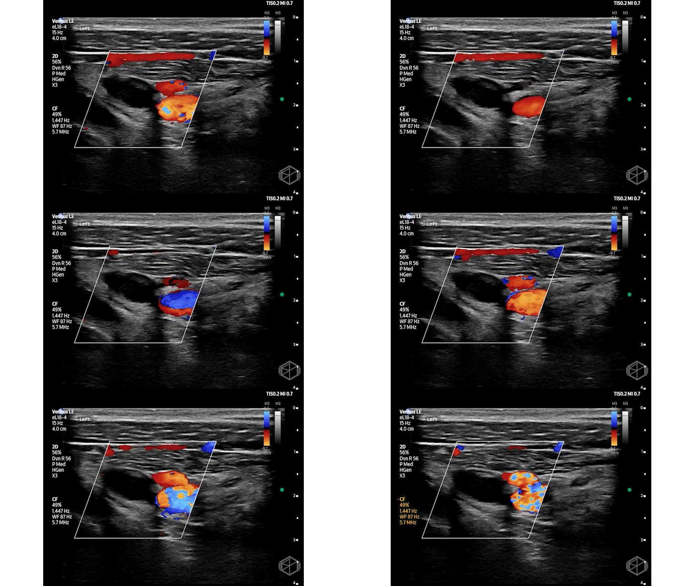
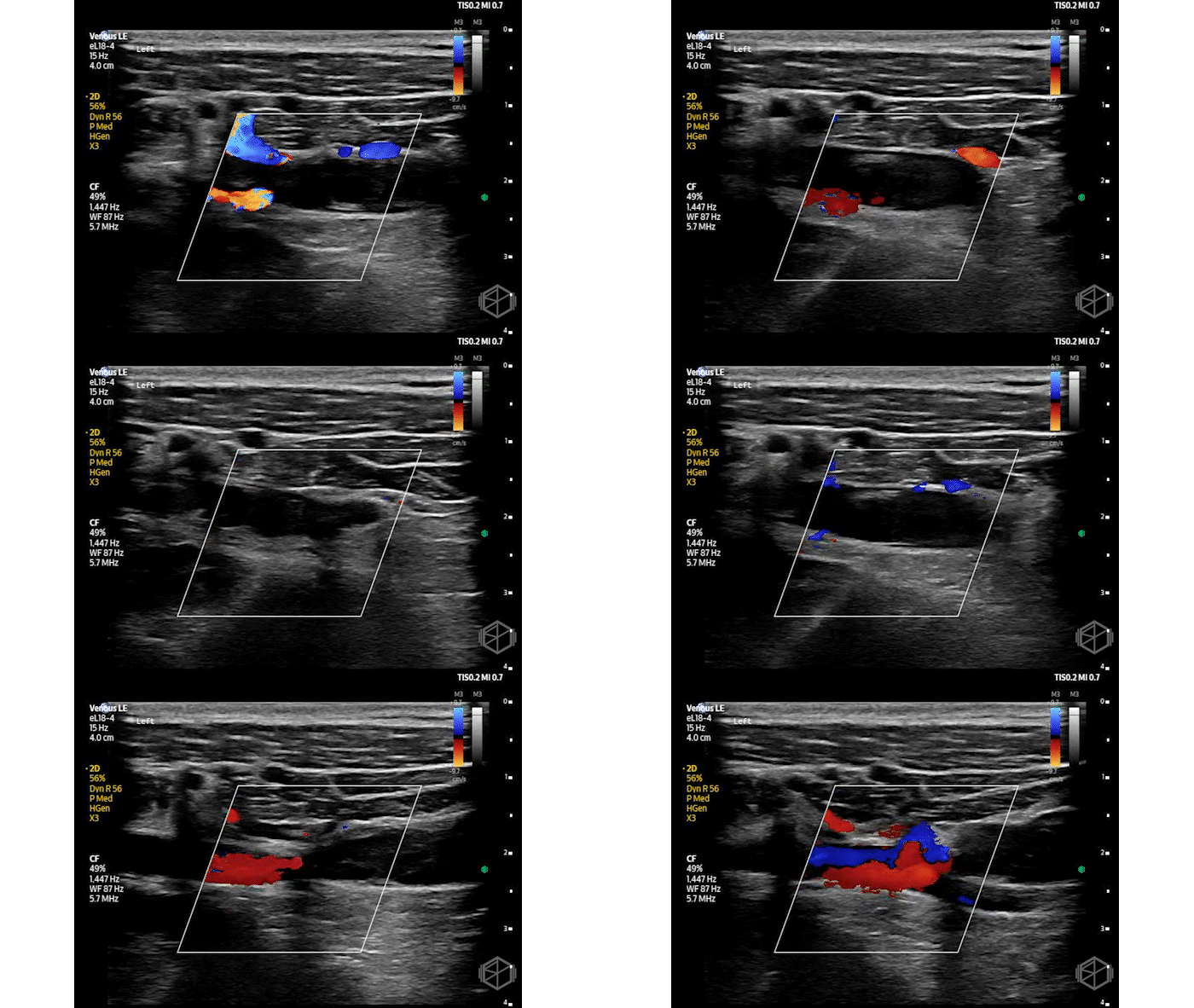
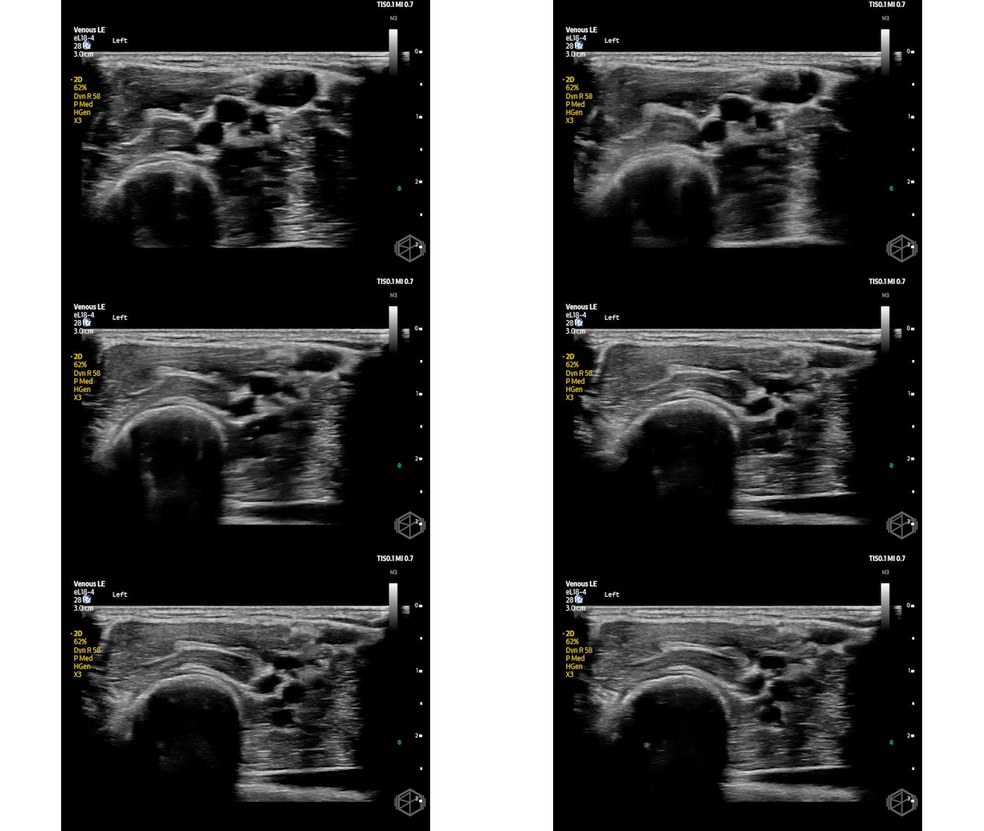
POCUS demonstrated an occlusive thrombus within the left subclavian vein. Radiology duplex ultrasound demonstrated the occlusive thrombosis involving the left subclavian and axillary veins.
The patient was admitted and started on a heparin infusion, followed by surgical thrombectomy the following day and then discharged on oral anticoagulation. There are plans for surgical resection of the left first rib.
Diagnosis: Acute left axillosubclavian deep vein thrombosis, concerning for Paget-Schroetter syndrome (PSS), also known as effort thrombosis.
Learning points:
Upper-extremity DVT (UEDVT) is uncommon, accounting for approximately 4–11% of all DVTs. UEDVT can be classified as:
PSS also known as effort thrombosis, is a form of primary UEDVT typically seen in young, otherwise healthy individuals. It is associated with repetitive upper-extremity activity and often occurs in the setting of venous thoracic outlet syndrome (VTOS). The underlying mechanism involves compression of the subclavian vein, typically between the clavicle and first rib, resulting in repetitive endothelial injury and thrombosis.
Identifying an axillosubclavian DVT in a young, otherwise healthy patient should prompt consideration of underlying venous thoracic outlet compression. Further evaluation with CT venography, MR venography, or catheter venography may be necessary to identify anatomical compression. Dynamic imaging with provocative arm positioning can help assess positional venous obstruction, although venous narrowing alone does not establish clinically significant VTOS. (📚 PMIDs: 25673095, 42276365)
As discussed in previous SonoProps, the subclavian vein presents a unique challenge during ultrasound evaluation because it cannot be completely compressed beneath the clavicle. Evaluation of the central subclavian vein relies more heavily on direct visualization and Doppler assessment, while compression ultrasound remains useful for evaluating the internal jugular, brachial, and accessible portions of the axillary veins.
Color Doppler may demonstrate absent or diminished flow, while spectral Doppler can reveal abnormal respiratory phasicity or loss of normal cardiac pulsatility, suggesting proximal venous obstruction.
A negative ultrasound does not reliably exclude subclavian thrombosis when clinical suspicion remains high. Additional imaging may be necessary to evaluate central venous obstruction or define the extent of thrombosis. (📚 PMIDs: 15914687, 32370975)
Effort thrombosis isn't limited to athletes or strenuous overhead activity. Musicians can develop it too. A published case report describes a violin player who developed subclavian vein thrombosis following a change in playing habits, a presentation the authors referred to as "bow syndrome." Repetitive positioning and movement of the upper extremity may contribute to venous compression even without strenuous exercise. (📚 PMID: 27823916)
Management of Paget-Schroetter syndrome may involve more than anticoagulation. Patients with acute effort thrombosis may benefit from early thrombus removal and, in selected cases, surgical thoracic outlet decompression.
A 2023 systematic review and meta-analysis involving 2,653 patients found that approximately 54% of patients treated with anticoagulation alone were symptom-free at follow-up, compared with 96% of patients treated with catheter-directed thrombolysis and first-rib resection. However, the certainty of comparative evidence was very low, and treatment decisions must be individualized. (📚 PMID: 37678659)
Long-term functional recovery is especially important in young patients with effort thrombosis. Persistent venous obstruction, recurrent thrombosis, and post-thrombotic symptoms may limit return to sports or other repetitive upper-extremity activities.
A systematic review of 123 reported cases of Paget-Schroetter syndrome in athletes found an average return-to-sport time of approximately 4.7 months. However, these findings are from athletic populations and should not be used to predict an individual patient's recovery. For a young musician, preservation of upper-extremity function may be particularly important. (📚 PMID: 32868012)
The next SonoProps goes to Dr. Mohsin Mohamedali!
An approximately 3-year-old female with no significant past medical history presented to the ED with left knee pain, swelling, and refusal to bear weight that began earlier that morning. Her parents reported no significant trauma, fever, or recent illness, although she had experienced a brief episode of diarrhea several days earlier. The hip was normal.
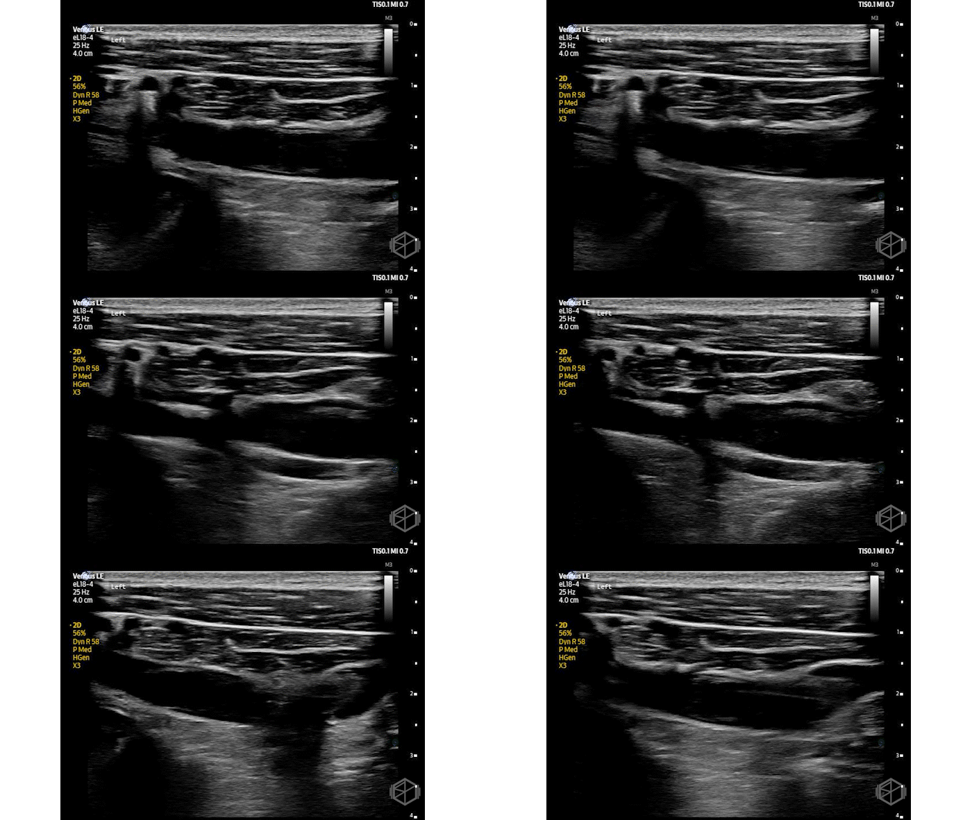
Plain radiographs also suggested the presence of a joint effusion. POCUS of the left knee demonstrated the following:
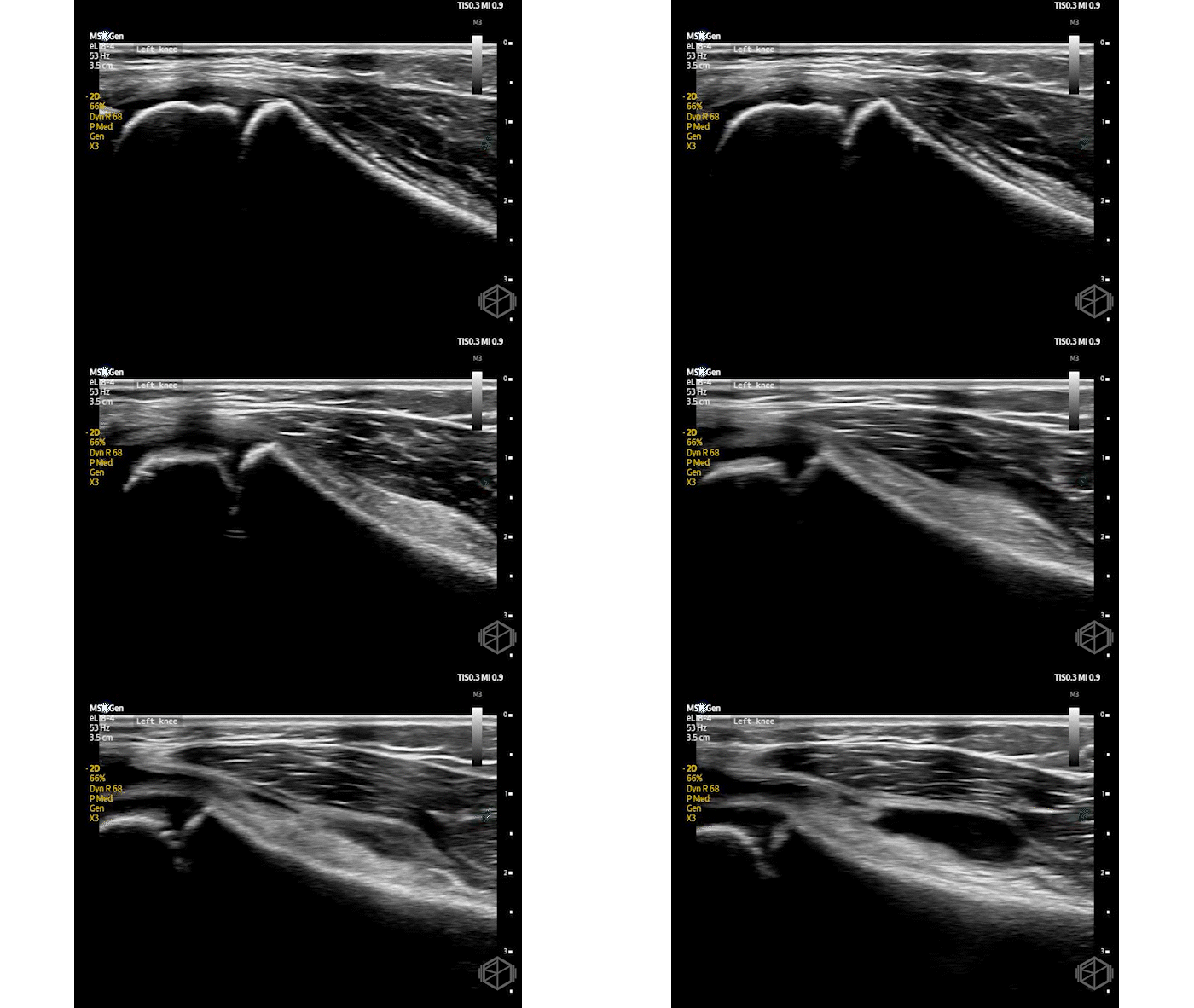
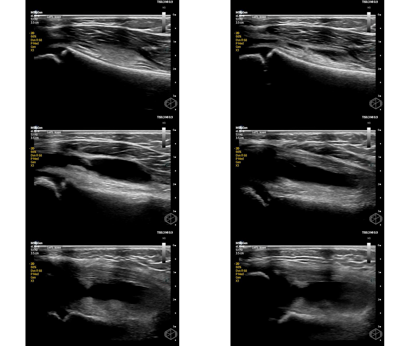
The clips above demonstrate a mildly complex left knee joint effusion. Laboratory studies revealed an ESR of 42 mm/hr and CRP of 47 mg/L.
Subsequent arthrocentesis demonstrated cloudy, bloody synovial fluid containing 103,320 nucleated cells/µL with 99% neutrophils, raising significant concern for septic arthritis.
The patient underwent operative irrigation and debridement, where purulent, bloody fluid was encountered. She was treated with antibiotics for presumed bacterial septic arthritis. Subsequent tissue cultures grew organisms that were interpreted as contaminants. Interestingly, Lyme serology returned strongly positive, with all 10 IgG immunoblot bands reactive, raising concern for Lyme arthritis.
Diagnosis: Acute inflammatory left knee effusion, concern for septic arthritis versus Lyme arthritis.
Learning Points:
POCUS is a valuable tool for evaluating pediatric patients with acute joint swelling or refusal to bear weight. Ultrasound can rapidly identify joint effusions and help distinguish intra-articular fluid from surrounding soft tissue swelling. It is helpful when physical examination is limited by pain or patient cooperation.
In a study involving 71 pediatric patients, knee radiographs demonstrated only 65% sensitivity and 84% specificity for identifying suprapatellar effusions, using assessments by experienced POCUS faculty as the reference standard. This highlights the limitations of radiographs for identifying joint fluid. (📚 PMID: 34542989)
The 2023 PIDS/IDSA guidelines recommend ultrasound when additional imaging is necessary to identify suspected joint effusions in children, particularly in joints that are more difficult to evaluate clinically. (📚 PMIDs: 37941444)
The suprapatellar recess is an important target when performing knee POCUS. A high-frequency linear transducer is typically placed longitudinally just superior to the patella to visualize the quadriceps tendon, suprapatellar recess, and distal femur.
Joint effusions typically appear as anechoic or hypoechoic fluid distending the suprapatellar recess, deep to the quadriceps tendon. Scanning in both longitudinal and transverse planes, including the medial and lateral parapatellar recesses, can help identify smaller effusions and distinguish intra-articular from extra-articular fluid collections.
Small effusions may be more apparent with gentle dynamic maneuvers or changes in knee positioning. A study comparing three ultrasound techniques found that quadriceps contraction identified more suprapatellar effusions than static scanning, although this maneuver may be difficult in younger children. (📚 PMIDs: 27812885, 32032167)
The sonographic appearance of a joint effusion can provide useful information, but cannot reliably distinguish septic arthritis from other inflammatory conditions. Simple effusions are typically anechoic, while complex effusions may demonstrate internal echoes, debris, or heterogeneous material.
A published case report described a toddler presenting with acute knee swelling and abnormal gait whose POCUS demonstrated complex, heterogeneous knee effusions. Further evaluation ultimately revealed juvenile idiopathic arthritis rather than bacterial septic arthritis. (📚 PMID: 30180103)
Although complex fluid may raise concern for an inflammatory, hemorrhagic, or infectious process, echogenicity alone cannot establish the etiology of the effusion.
POCUS can also facilitate arthrocentesis by identifying the location, size, and accessibility of a joint effusion. It can help select an appropriate needle entry site, visualize surrounding structures, and guide needle advancement into the joint space.
A pediatric emergency medicine case series demonstrated successful POCUS-guided identification and aspiration of effusions involving the knee, hip, shoulder, and elbow. (📚 PMID: 34318953)
Studies evaluating ultrasound-guided arthrocentesis suggest potential improvements in procedural accuracy and successful aspiration compared with landmark-guided techniques. Pediatric comparative evidence is limited, and ultrasound guidance may be particularly useful when effusions are small or difficult to palpate. (📚 PMIDs: 22103390, 41169402)
A joint effusion on POCUS does not necessarily mean bacterial septic arthritis. Septic arthritis, Lyme arthritis, reactive arthritis, and juvenile idiopathic arthritis can produce similar sonographic findings, including synovial thickening, effusions, and internal debris.
In Lyme-endemic regions, Lyme arthritis is an important consideration in children presenting with atraumatic knee swelling. Synovial fluid WBC counts can be markedly elevated in both bacterial septic arthritis and Lyme arthritis, making the distinction challenging.
Our patient's synovial fluid contained more than 100,000 nucleated cells/µL with 99% neutrophils, initially raising significant concern for bacterial septic arthritis. However, her strongly positive Lyme serology introduced an alternative explanation for the inflammatory effusion.
POCUS identifies and characterizes the effusion, but laboratory testing, arthrocentesis, and clinical evaluation remain essential to determining the underlying diagnosis. (📚 PMIDs: 24365728, 29669751, 30180103)
