
October SonoProps
Welcome to our October SonoProps!
The first SonoProps for this month goes to Dr. Yianni Flouskakos and Dr. Chirag Parikh.
A teenage male was brought to the ED following a witnessed cardiac arrest. Upon EMS arrival, the patient was found to be in asystole and ACLS was initiated. He received epinephrine and subsequently developed ventricular fibrillation, requiring defibrillation. His rhythm returned to asystole before progressing to torsades de pointes, for which he received 2 g of magnesium. ROSC was eventually achieved, and the patient was intubated prior to ED arrival.
POCUS demonstrated the following findings:
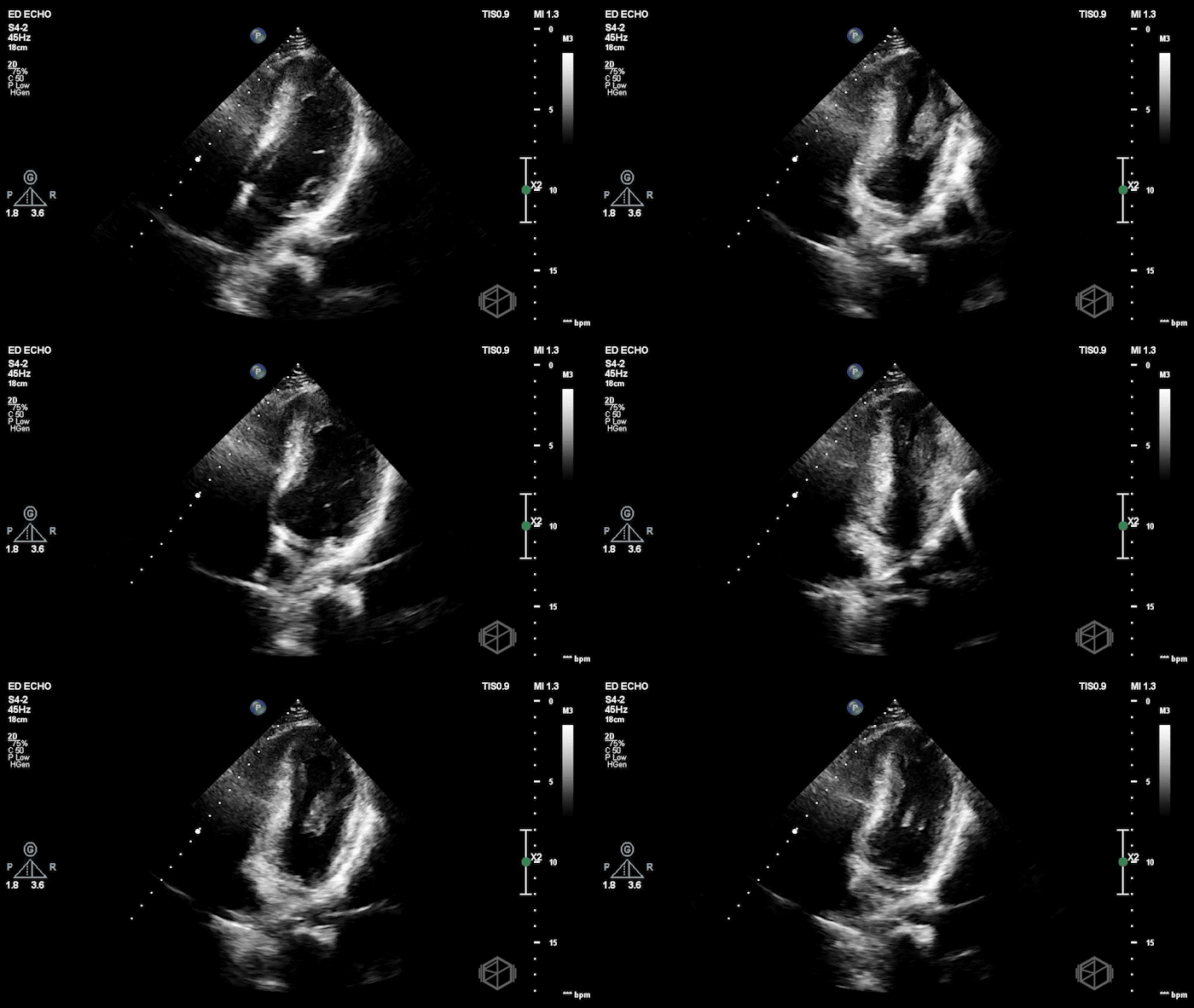
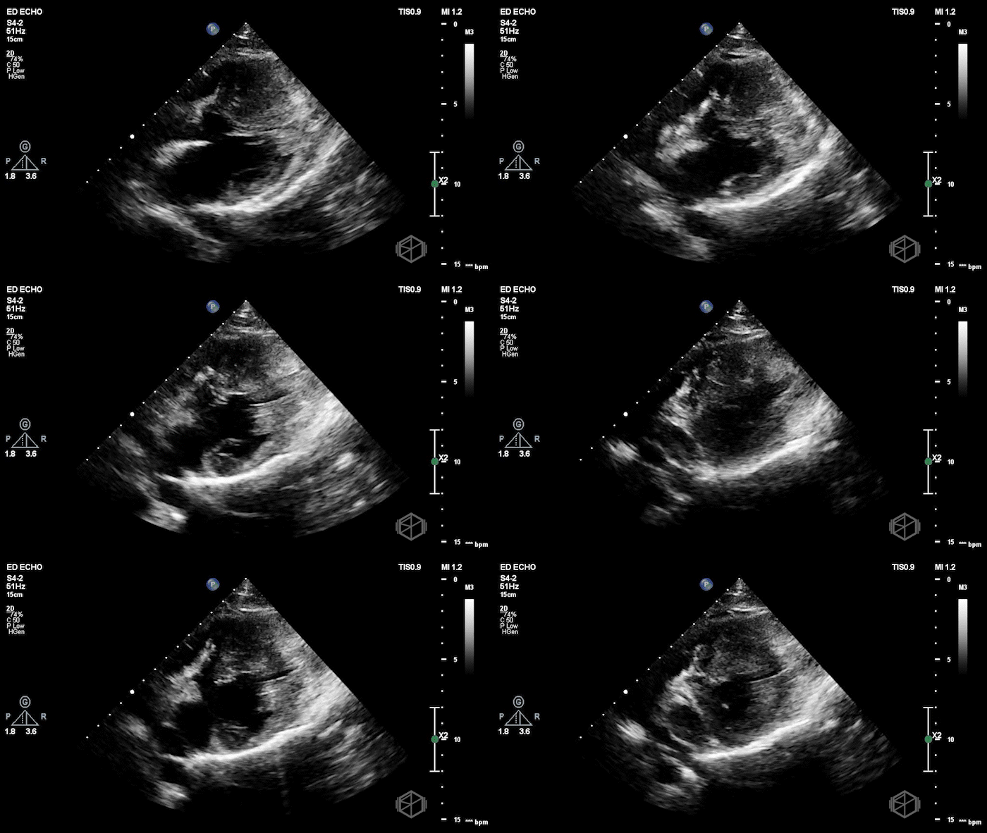
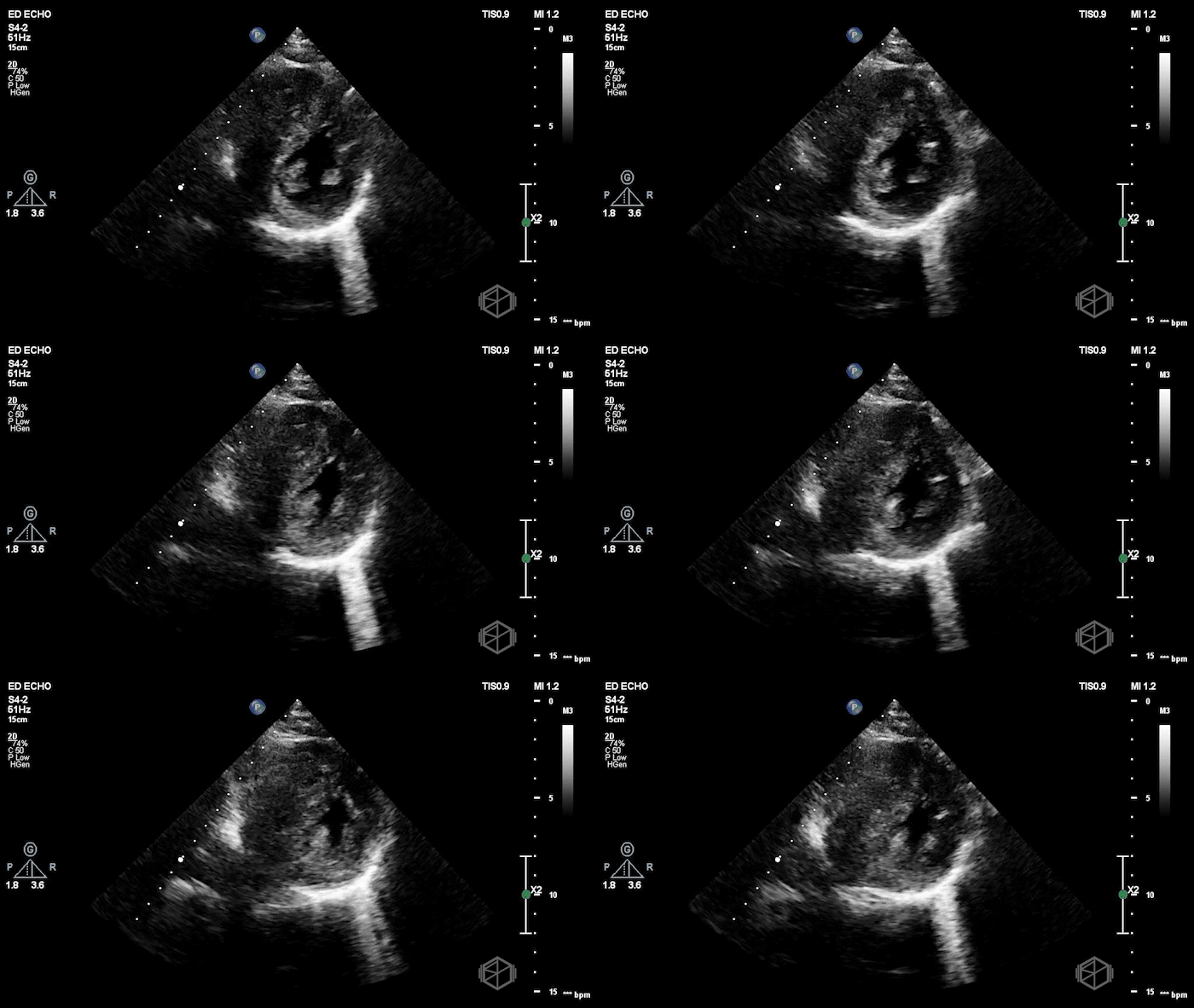
The POCUS shows a hyperdynamic left ventricle with severe asymmetric left ventricular hypertrophy, most pronounced in the interventricular septum, anterior wall, and apex, with relatively minimal hypertrophy of the inferior wall. There is also systolic anterior motion of the mitral valve, although it was difficult to determine if there was left ventricular outflow tract obstruction. It was later discovered that a patient had a a known history of hypertrophic cardiomyopathy (HCM), on atenolol.
Diagnosis: HCM with systolic anterior motion (SAM) of the mitral valve, presenting with Vfib arrest
Learning Points:
HCM is characterized by unexplained left ventricular hypertrophy, most commonly involving the interventricular septum. Although asymmetric septal hypertrophy is the classic presentation, hypertrophy can also involve the anterior wall, apex, or other LV segments. POCUS may demonstrate disproportionate wall thickening, a small LV cavity, and preserved or hyperdynamic systolic function.
In adults, a maximal LV wall thickness ≥15 mm, not explained by other conditions, generally supports the diagnosis of HCM. Patients with a family history of HCM or a pathogenic genetic variant may meet diagnostic criteria at lesser degrees of hypertrophy. Diagnostic criteria differ in children. POCUS findings should prompt comprehensive cardiac evaluation when HCM is newly suspected. (📚 PMID: 28912181)
SAM of the mitral valve occurs when the mitral valve moves toward the interventricular septum during systole. It is best visualized in the parasternal long-axis view and can contribute to dynamic left ventricular outflow tract (LVOT) obstruction and mitral regurgitation.
SAM results from altered ventricular geometry, abnormalities of the mitral valve apparatus, and blood-flow-related drag forces. The mitral leaflet may contact the septum during systole, narrowing the LVOT. SAM may also cause mitral regurgitation due to incomplete closure of the mitral valve leaflets. (📚 PMID: 35813751)
SAM does not always result in significant LVOT obstruction. Obstruction is dynamic and influenced by preload, afterload, contractility, and heart rate. Decreased preload, decreased afterload, and increased contractility can worsen obstruction, while patients may have minimal gradients at rest. The presence of SAM alone on 2D POCUS cannot establish the severity of LVOT obstruction.
Advanced echo: A peak instantaneous LVOT gradient ≥30 mmHg defines obstruction, while ≥50 mmHg is generally considered the threshold for significant obstruction when considering advanced therapies. Color Doppler may identify turbulent flow, but continuous-wave Doppler is required to quantify the gradient. (📚 PMIDs: 38718139, 35813751)
HCM is an important cause of sudden cardiac death in adolescents and young adults, typically due to malignant ventricular arrhythmias such as VT or VF. Myocardial disarray, fibrosis, and ischemia contribute to electrical instability, and sudden cardiac arrest may occur even in patients without significant LVOT obstruction.
Risk factors include prior cardiac arrest, unexplained syncope, family history of sudden cardiac death, massive LV hypertrophy, and nonsustained VT. Survivors of cardiac arrest attributable to VT/VF generally have an indication for ICD placement for secondary prevention. (📚 PMIDs: 34636345, 38718139)
Recognizing HCM and SAM on POCUS can influence management of hypotension and shock. Dynamic LVOT obstruction can worsen with dehydration, vasodilation, or medications that increase cardiac contractility. Recognizing this physiology on POCUS may change how hypotension is treated. SAM alone does not prove that LVOT obstruction is causing the patient's shock.
Reduced preload or afterload and increased contractility may narrow the LVOT further and reduce cardiac output.
In hypotension attributable to obstructive HCM, treatment may include correcting hypovolemia when present and using vasoconstrictors without significant inotropic effects, such as phenylephrine. Post-cardiac arrest hypotension is often multifactorial, so SAM should prompt additional evaluation rather than automatically determine treatment. (📚 PMIDs: 38718139, 35813751)
The second SonoProps goes to Dr. Rachel Ariz!
An approximately 70-year-old female with recently diagnosed stage IV colon cancer, currently receiving oral chemotherapy, presented to the ED with sudden-onset diffuse abdominal pain beginning approximately one hour prior to arrival. The pain was severe, persistent, and generalized, without associated vomiting, fever, diarrhea, or gastrointestinal bleeding.
Her medical history was also significant for a recent hospitalization for neutropenic fever secondary to enteritis and a Pseudomonas mediport infection. She was anticoagulated with Eliquis for a recent right internal jugular DVT.
Given her acute abdominal pain and significant medical history, bedside POCUS was performed, revealing the following:
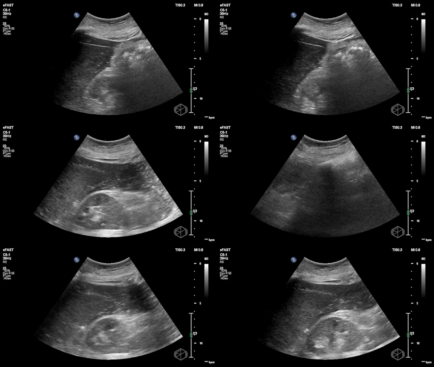
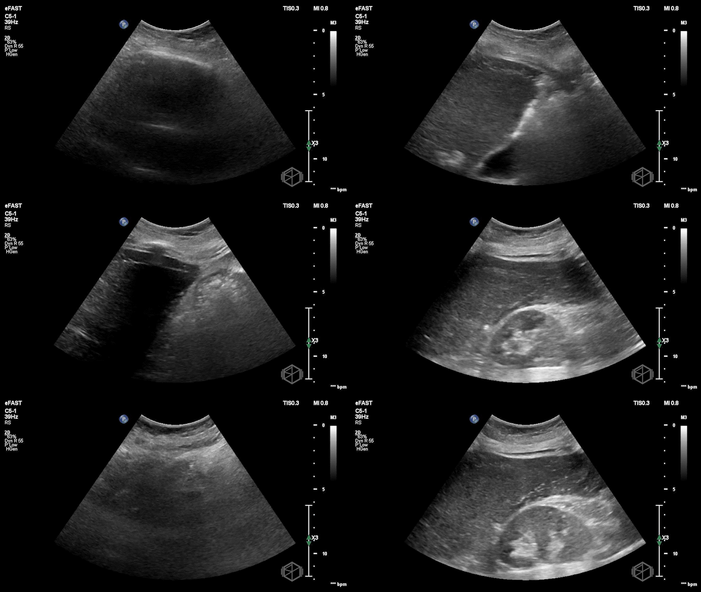
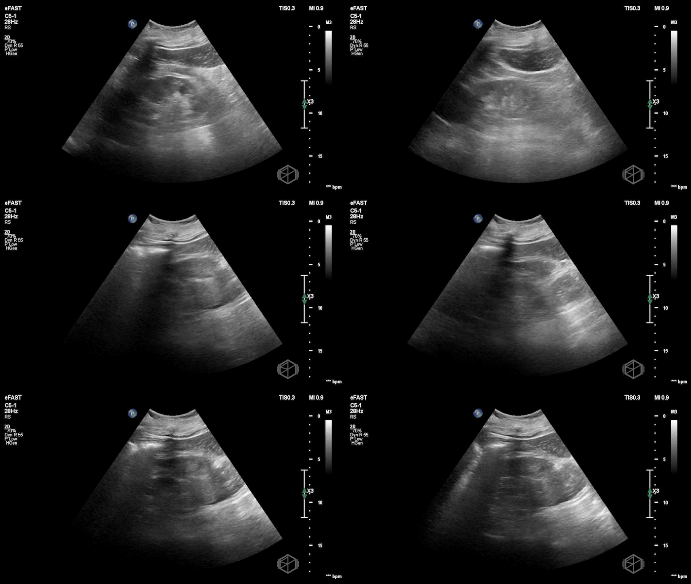
POCUS demonstrated complex free fluid in both the right and left upper quadrants. The fluid appeared diffusely echogenic and relatively uniform, without prominent septations or layering.
Subsequent CT imaging demonstrated moderate pneumoperitoneum with free fluid, predominantly in the upper and right abdomen, concerning for perforation of the ascending or proximal transverse colon.
Diagnosis: Complex intra-abdominal free fluid with pneumoperitoneum concerning for colonic perforation in a patient with metastatic colon cancer
Learning points:
Complex free fluid does not always have to look complex, as mentioned in prior learning points. While complex fluid often contains septations, layering, or heterogeneous debris, it may also appear relatively homogeneous with subtle, diffuse low-level internal echoes. Careful evaluation of fluid echogenicity is important, as hemorrhage, infection, and inflammatory exudate may resemble simple ascites. (📚 PMIDs: 28567101, 12740467)
Complex free fluid in acute abdominal pain should increase concern for bowel perforation and peritonitis. Leakage of intestinal contents and inflammatory exudate can produce echogenic or "dirty" intraperitoneal fluid, sometimes before free air is recognized on POCUS. Although nonspecific, this finding should prompt consideration of hollow-viscus perforation, especially in patients with sudden severe abdominal pain. (📚 PMID: 27274522)
POCUS can potentially help identify pneumoperitoneum. The enhanced peritoneal stripe sign (EPSS) appears as abnormal brightening of the anterior peritoneal line, potentially accompanied by reverberation artifacts, dirty shadowing, or ring-down artifacts. The right upper quadrant, particularly the prehepatic region beneath the anterior abdominal wall, is a useful location to evaluate for free intraperitoneal air. A high-frequency linear probe may improve visualization. (📚 PMIDs: 10541100, 23902744)
The absence of pneumoperitoneum on POCUS does not exclude bowel perforation. While earlier studies demonstrated promising diagnostic performance, a 2025 study evaluating emergency physicians' interpretation of ultrasound clips during controlled peritoneal insufflation reported 66% sensitivity and 85% specificity for pneumoperitoneum, with sensitivity increasing as free-air volume increased. A negative examination should not delay CT or surgical evaluation when clinical suspicion remains high. (📚 PMIDs: 17347768, 39862481)
Do not anchor on malignant ascites in a patient with known intra-abdominal cancer. Although peritoneal carcinomatosis can produce complex ascites, sudden severe abdominal pain should prompt consideration of perforation, obstruction, hemorrhage, or secondary peritonitis. Fluid echogenicity alone cannot reliably distinguish malignant, infected, or hemorrhagic ascites. Clinical context and further imaging is incredibly useful. (📚 PMIDs: 18060209, 110040)
