
September SonoProps
September brings us two great examples of how POCUS can guide both diagnosis and procedural care in the ED.
Our first September SonoProps goes to Drs. Siri Tummala and Mohsinali Mohamedali. They were working with our fellow Dr. Brian Tang and obtained this great scan.
They had a woman in her mid-40’s with a history of tobacco use who presented to the ED with acute onset shortness of breath and cough.
Dr. Tang performed the following scan that showed:
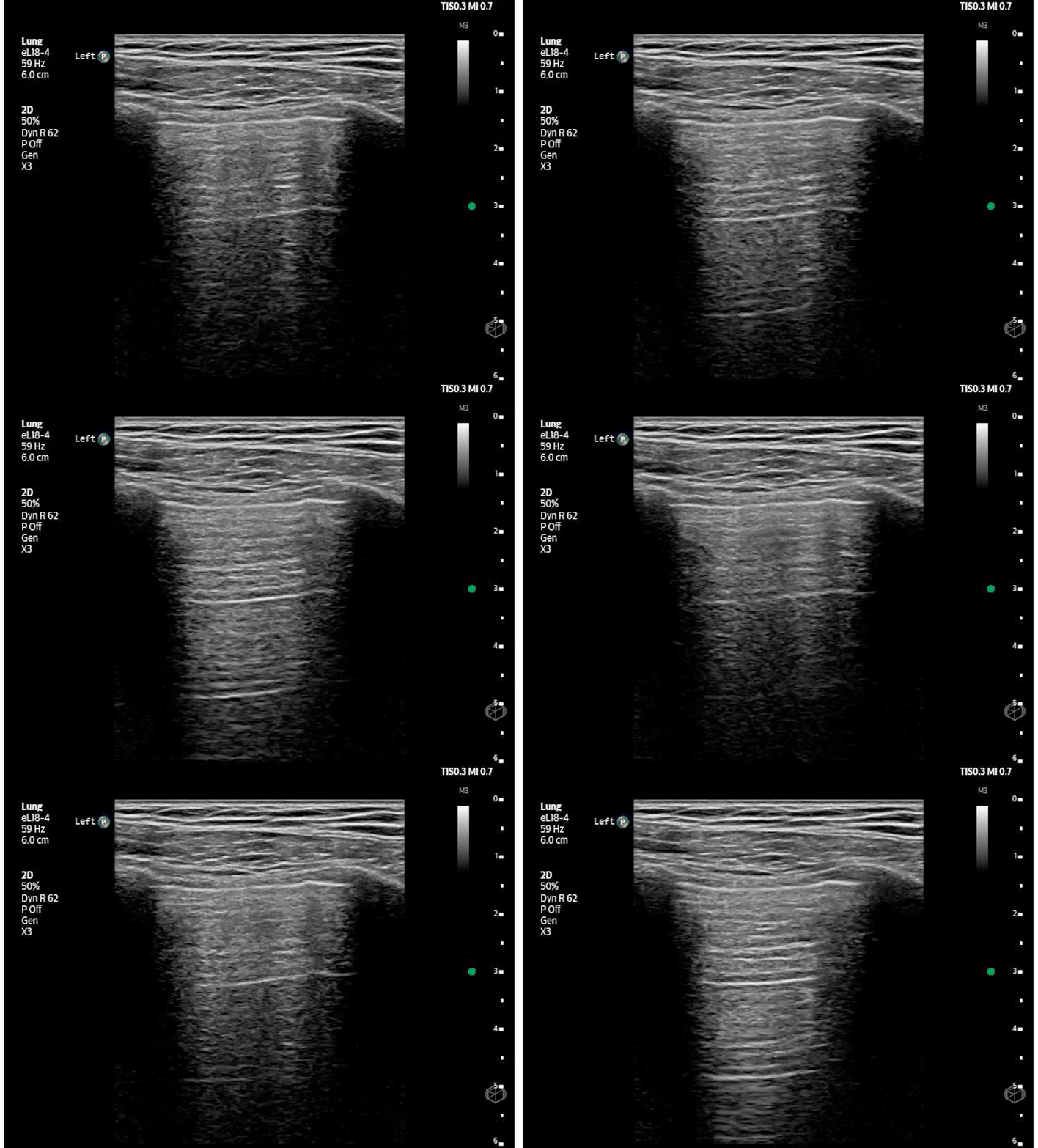
Lung ultrasound demonstrated absent lung sliding on the left, raising concern for pneumothorax.
Given that the patient was stable, chest radiography was obtained and confirmed a large left-sided pneumothorax. The team then proceeded with pigtail thoracostomy. Drs. Mohamedali, Tummala, and Tang performed the following block to facilitate pigtail placement.
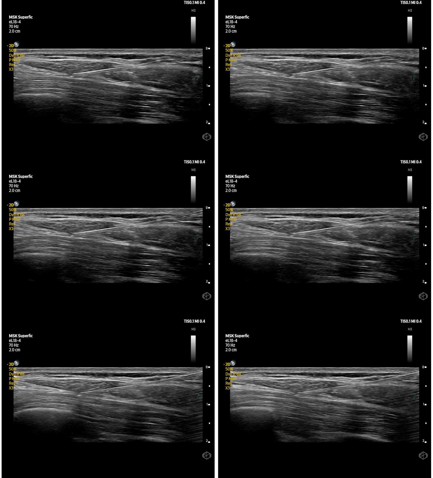
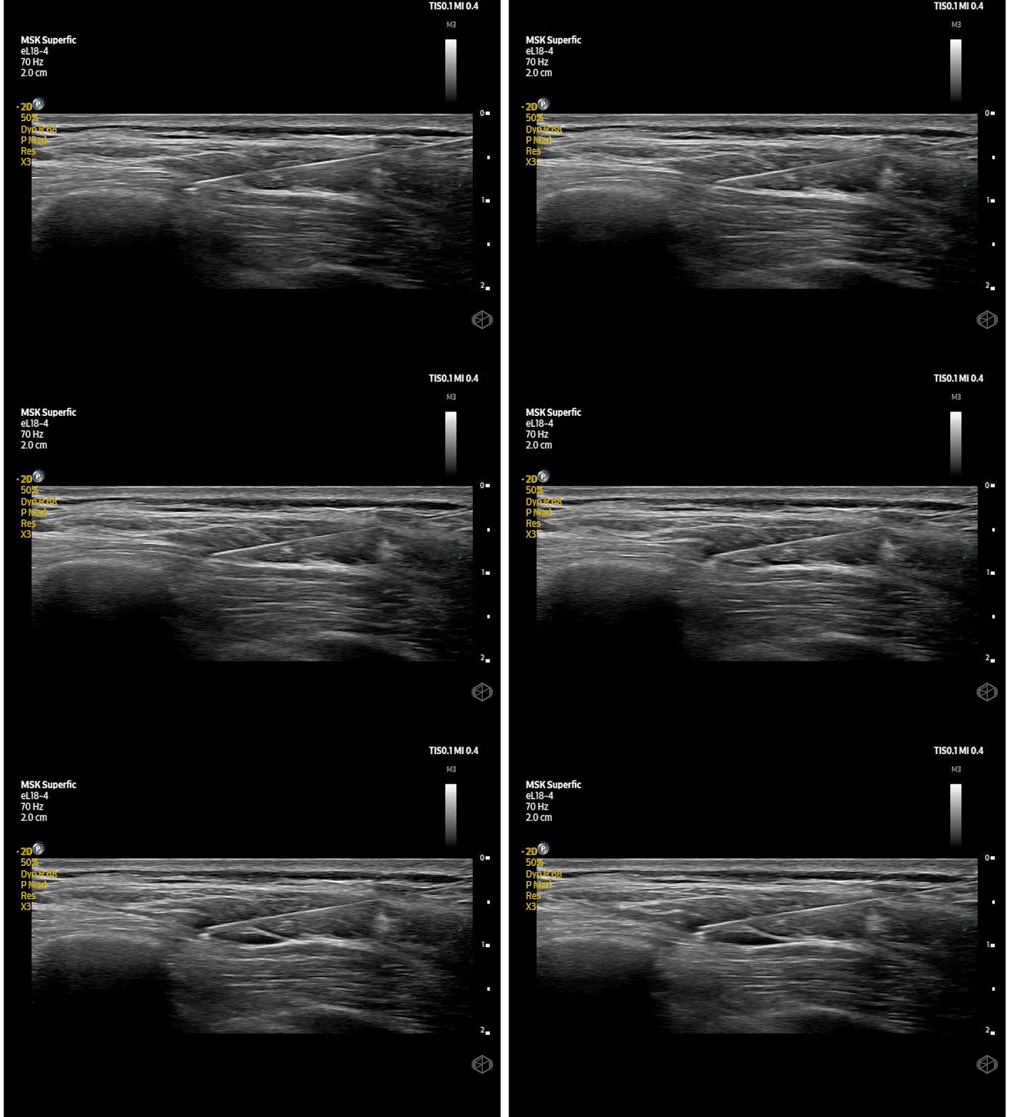
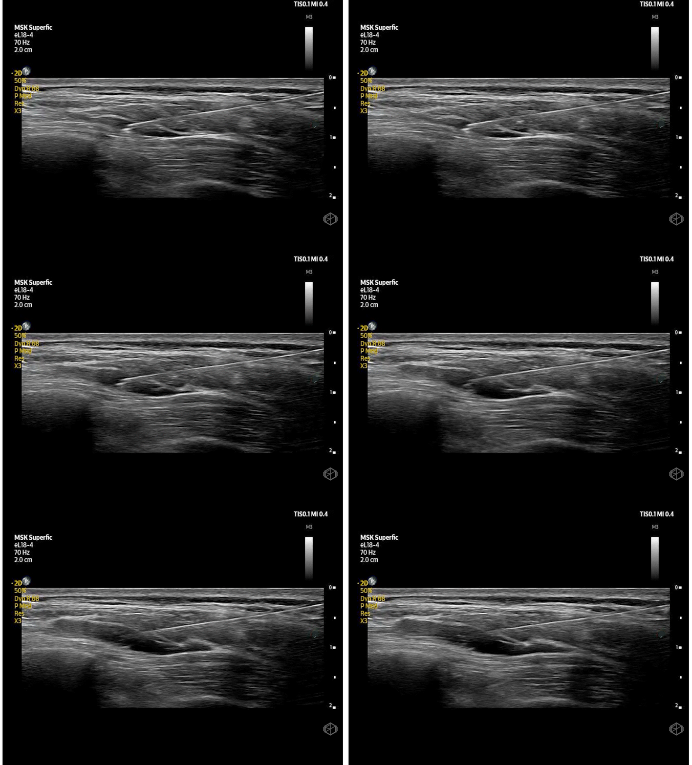
The patient tolerated the procedure well overall but required additional local anesthesia during tract dilation, potentially due to incomplete anesthesia of the deeper intercostal structures and parietal pleura.
Diagnosis: Pneumothorax, with pigtail placement facilitated by a Serratus Anterior Plane Block
Learning Points:
SAPB can be a useful option for chest wall analgesia and painful procedures such as pigtail thoracostomy. The block targets the lateral cutaneous branches of the intercostal nerves and can provide analgesia over the lateral chest wall. Case reports and small series describe its use before chest tube and pigtail placement in the ED, with some patients achieving nearly complete or complete procedural anesthesia.
SAPB can anesthetize the overlying skin, but its effects on deeper structures, particularly the intercostal nerves and parietal pleura, are variable. Assess the block before starting the procedure and provide supplemental local anesthetic infiltration along the planned insertion tract when needed.
SAPB primarily covers the anterolateral chest wall, while posterior coverage may be less reliable. An erector spinae plane block may provide broader posterior chest wall analgesia, particularly for posterior rib fractures.
(📚 PMIDs: 27440171, 39903631, 41567479, 40379486)
Start by identifying the anatomy. Place a high-frequency linear probe over the lateral chest wall around the fourth or fifth rib. Identify the latissimus dorsi, serratus anterior, rib, and pleural line. The superficial approach places anesthetic between latissimus dorsi and serratus anterior, while the deep approach targets the plane between serratus anterior and the rib. (📚 PMID: 39903631, 27440171)
Use hydrodissection to confirm that you are in the right plane. Once the needle tip is in position, inject a small amount of fluid and watch for separation of the fascial layers. The goal is to see fluid track along the plane rather than expand the muscle itself. (📚 PMID: 39903631, 41567479)
Keep the needle tip in view. The superficial approach to the serratus anterior plane block is relatively safer than the deep approach, but the pleura is still nearby. An in-plane approach helps keep the needle visible as it advances toward the target. The rib can also serve as a useful landmark and backstop, particularly with the deep approach. Pneumothorax has been reported as a complication, so continuous needle-tip visualization matters.
(📚 PMID: 32154513, 39903631)Watch the spread. This is a fascial plane block, so the local anesthetic needs to spread along the plane. If you see poor spread or intramuscular injection, reposition the needle before continuing.
(📚 PMID: 27440171, 27051373)
The next SonoProps goes to Dr. Ahmer Salman.
Dr. Salman evaluated a 65-year-old male with a history of alcoholic cirrhosis complicated by ascites who presented to the ED for evaluation of an elevated white blood cell count. The patient had recently undergone multiple abdominal procedures, including paracentesis and ERCP. Despite his abnormal laboratory findings, he reported feeling well and had no acute complaints.
He performed a scan that showed:
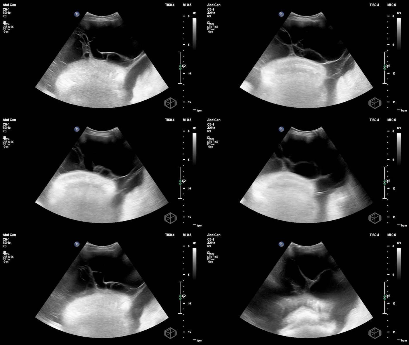
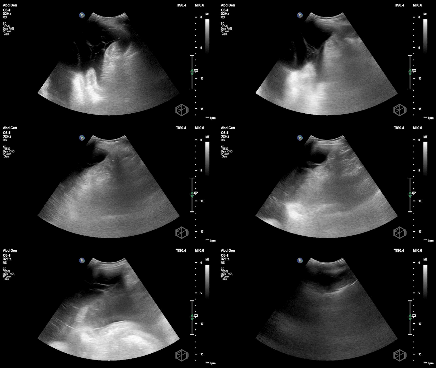
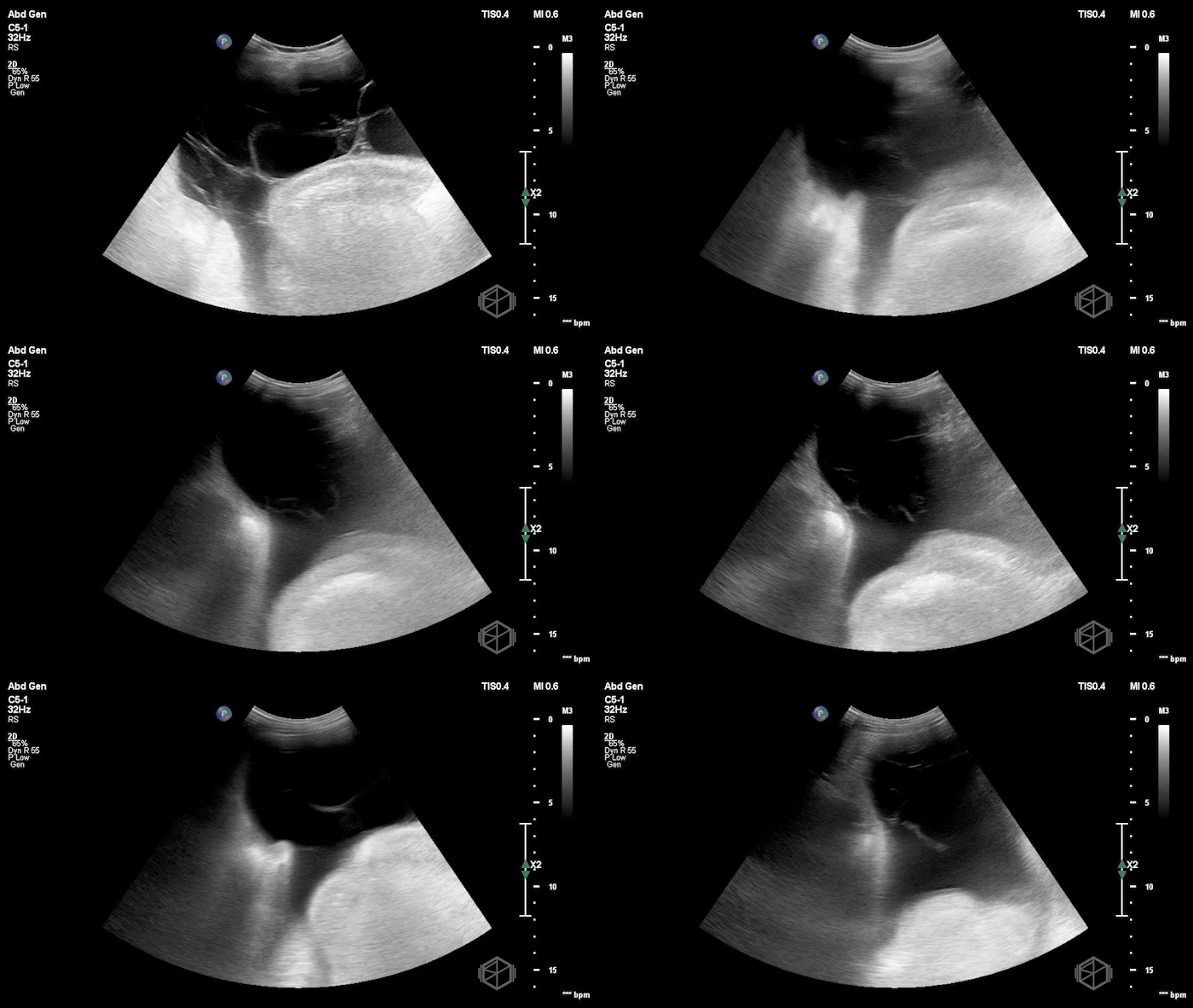
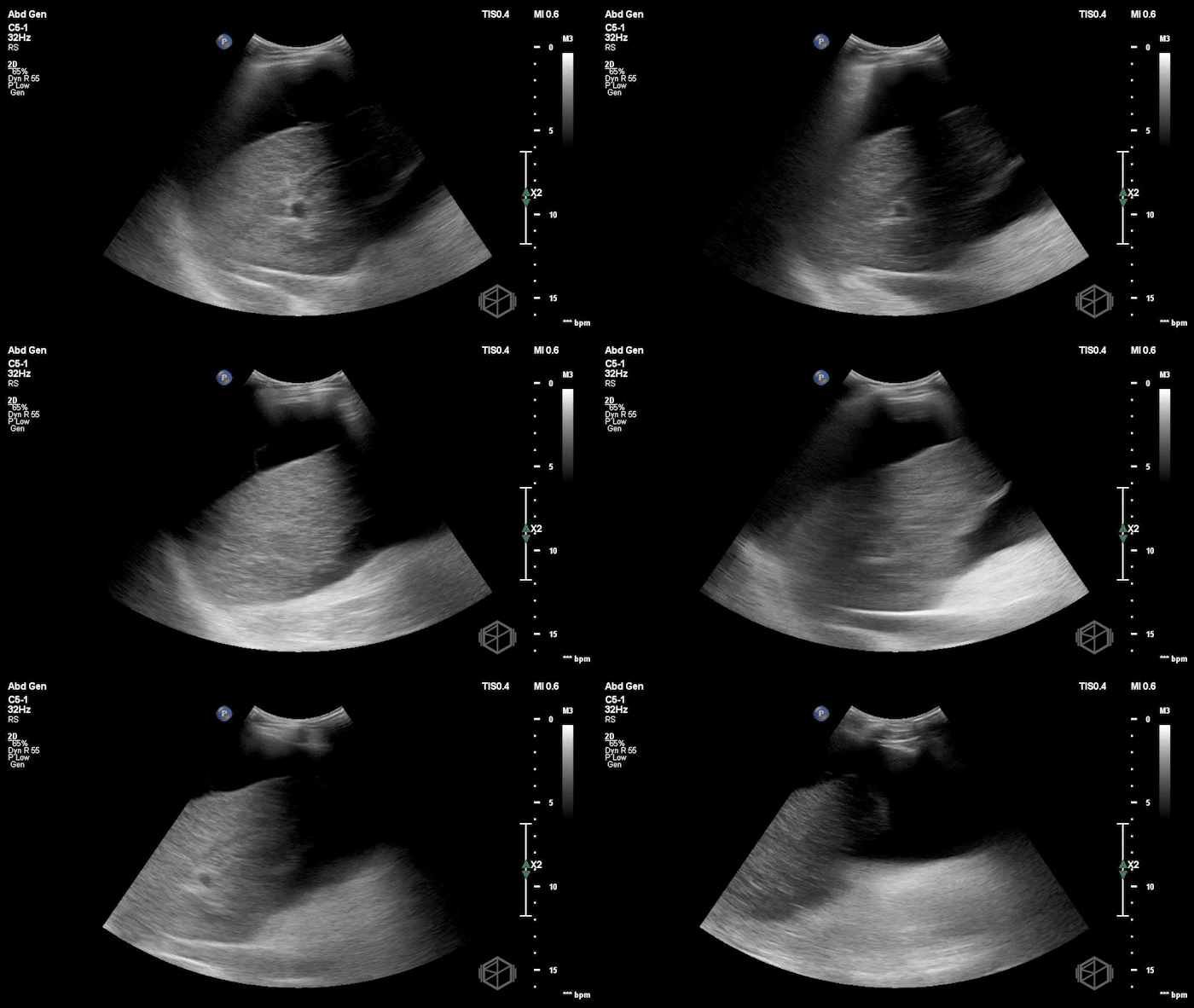
The ultrasound clips demonstrated complex ascites with internal septations. Dr. Salman performed an ultrasound-guided paracentesis, removing approximately 2 L of fluid. Fluid analysis revealed 3,884 nucleated cells/µL with 86% neutrophils, corresponding to an absolute PMN count of approximately 3,340 cells/µL. Even after correction for the bloody fluid, the PMN count remained significantly elevated, consistent with spontaneous bacterial peritonitis (SBP).
Diagnosis: Complex Ascites, SBP
Learning Points:
SBP can present without obvious abdominal symptoms. Patients with cirrhosis and ascites may have bacterial peritonitis despite a relatively reassuring clinical presentation. An elevated white blood cell count or other unexplained clinical deterioration should prompt consideration of infection. Ascitic fluid analysis is necessary to establish the diagnosis of SBP. (📚 PMID: 23473819)
Simple ascites is typically anechoic, while complex ascites may contain particulate debris, low-level echoes, layering, septations, or loculations. POCUS can help characterize this complexity, which may be less apparent on CT. These findings describe complex fluid rather than a specific diagnosis. Septations and debris have been described with infected ascites, but POCUS alone cannot reliably determine the cause. Diagnostic paracentesis should be performed when clinically indicated and feasible. (📚 PMID: 28567101, 110040, 33942342)
POCUS should evaluate the location and amount of fluid, size of the collection, abdominal wall thickness, adjacent organs, and anticipated needle trajectory. The planned site should also be evaluated in multiple planes before puncture. This becomes especially important when the fluid is septated, loculated, or otherwise difficult to access.
(📚 PMID: 30604780)POCUS can significantly change where a paracentesis is performed. In one prospective study, ultrasound changed the planned needle insertion site by ≥5 cm in 69% of procedures. The ultrasound-selected pocket was significantly deeper (5.4 vs 3.0 cm), and POCUS led clinicians to abort 13% of planned procedures because an adequate volume of ascites could not be safely identified. (📚 PMID: 34346007)
Use Doppler to make paracentesis safer. A fluid pocket can look ideal on grayscale while an abdominal wall vessel crosses the intended needle path. The Society of Hospital Medicine recommends color-flow Doppler evaluation along the anticipated needle trajectory to identify and avoid abdominal wall vessels. Injury to vessels such as the inferior epigastric artery can result in significant hemorrhage, abdominal wall hematoma, or pseudoaneurysm.
(📚 PMID: 30604780, 23546486, 9737471)Consider real-time ultrasound guidance for more difficult paracenteses. Static ultrasound marking is appropriate for many procedures, but real-time needle guidance should be considered when the fluid collection is small or difficult to access. A septated or loculated collection may be one situation where keeping the needle and target under continuous visualization is useful rather than simply marking a site and putting the probe down.
(📚 PMID: 30604780)
