
August SonoProps
Our first SonoProps goes to Dr. Marina Frayberg and Dr. Michael Gorynski!
A mid 60’s male with a recent complicated hospitalization for GI bleed presented to the ED with new left-sided neck pain and swelling. During his recent admission, he had undergone an uncomplicated left internal jugular central line placement for resuscitation that was removed without issue prior to his discharge.
Given the location of his symptoms and recent vascular access, POCUS of the neck was performed by Drs. Frayberg and Gorynski:
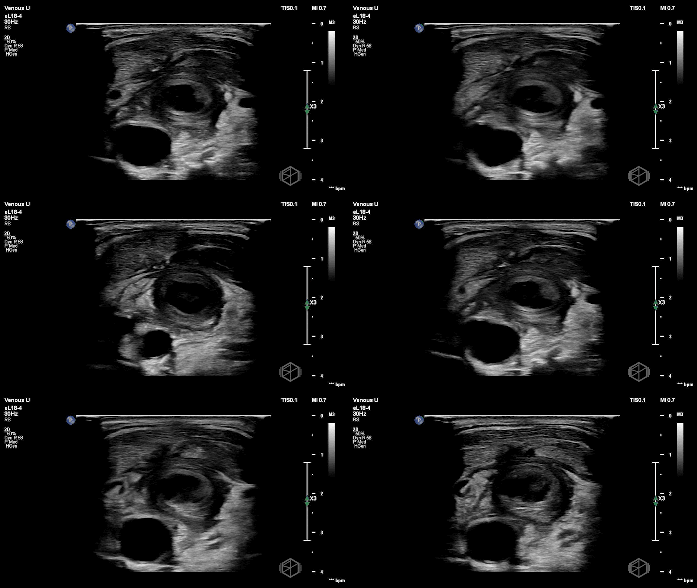
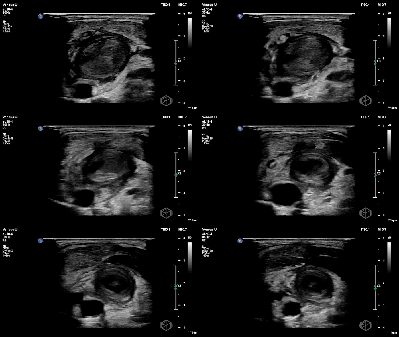
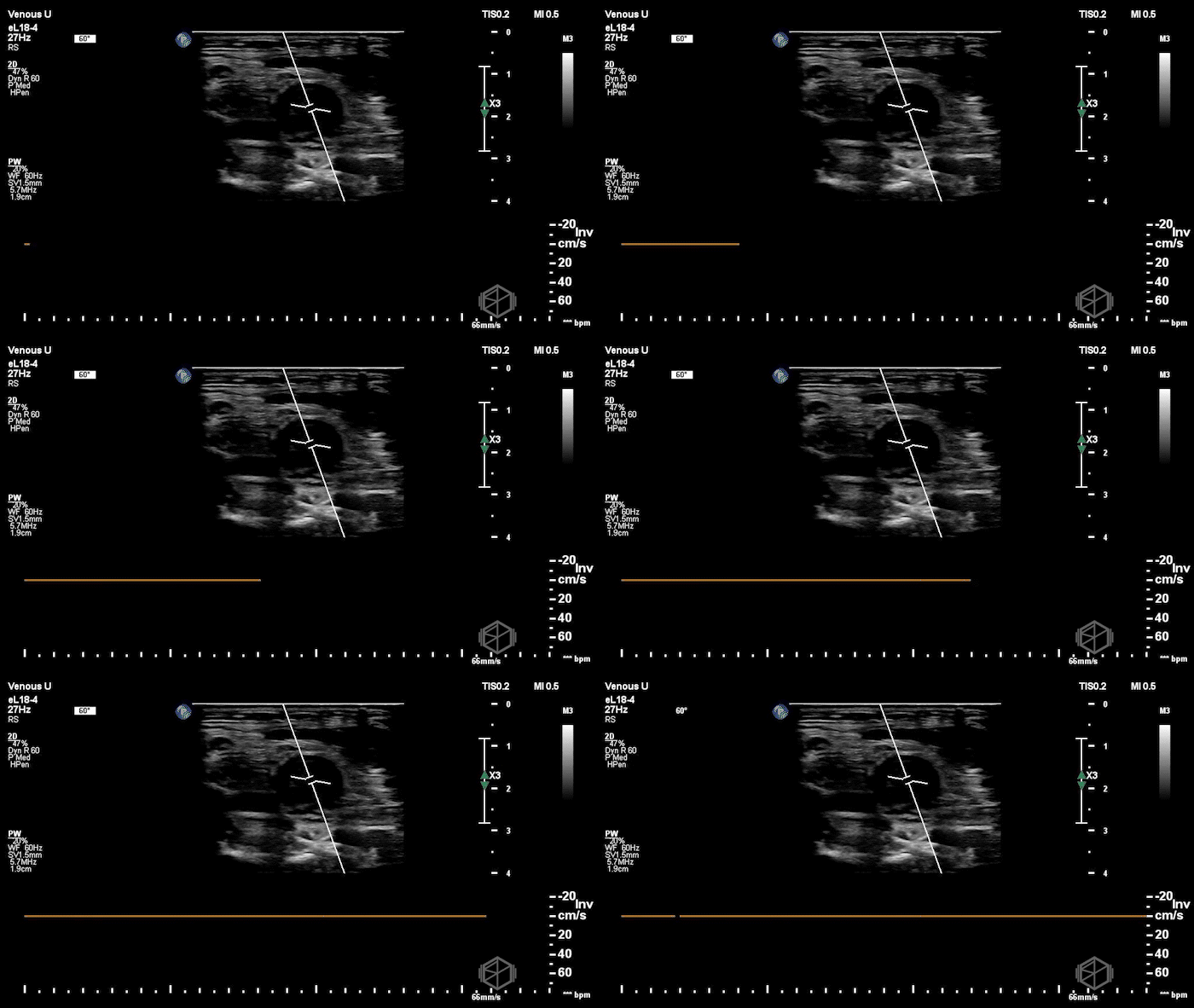
The left internal jugular vein contained extensive thrombus and was noncompressible, with surrounding inflammatory changes concerning for thrombophlebitis.
Vascular surgery was consulted and recommended additional imaging. Radiology ultrasound confirmed expansile occlusive thrombosis of the left internal jugular vein with surrounding soft-tissue edema and no drainable collection. CT of the neck demonstrated extensive left IJ thrombophlebitis with thrombus extending cranially into the distal left sigmoid sinus. The patient was started on heparin and antibiotics and admitted to Medicine for further management and monitoring given his recent GI bleed. ID considered Lemierre syndrome but favored catheter-associated IJ thrombosis with thrombophlebitis.
Diagnosis: Likely catheter-associated left internal jugular vein thrombosis with thrombophlebitis and extension into the distal sigmoid sinus
Learning Points:
Recent IJ access + new unilateral neck pain or swelling should raise concern for IJ thrombosis. Internal jugular vein thrombosis can present with neck pain, swelling, erythema, or tenderness. Central line placement is a well-described acquired risk factor. An uncomplicated insertion does not exclude a subsequent catheter-related thrombus. (📚 PMID: 33672254)
Catheter-associated thrombosis is not uncommon and may be asymptomatic. In one prospective study of critically ill adults who underwent duplex ultrasound after CVC removal, catheter-related DVT was found in 26% of the evaluated catheters; 70% of the identified thrombi involved the internal jugular vein. This was a selected ICU population, so the number should not be interpreted as the incidence for all IJ lines, but it highlights that thrombosis can occur even without an obvious complication during insertion. (📚 PMID: 28839334)
Compression is the key bedside maneuver. A normal IJ should collapse with gentle probe pressure. A persistently noncompressible vein, especially with visible intraluminal thrombus, should immediately raise concern for thrombosis. A systematic review of upper-extremity DVT found duplex ultrasound to have a pooled sensitivity of approximately 87% and specificity of 85%, although performance varies with the vessel being examined and study technique. (📚 PMIDs: 33672254, 32511715)
POCUS may not be able to determine how far the clot goes. The accessible portion of the IJ is easy to evaluate, but cranial extension toward the skull base and cerebral venous sinuses is much harder to assess. In this case, CT showed extension into the sigmoid sinus. Additional imaging may be needed to define the extent of thrombosis. (📚 PMID: 33672254)
Pulmonary embolism is considered a potential complication of IJ thrombosis, but the risk from an isolated IJ thrombus is poorly defined. A systematic review found surprisingly little direct evidence linking isolated IJVT to clinically overt PE: of 274 papers screened, only two described IJ thrombosis preceding PE in a way that suggested causality, and both were confounded. The review also found no clear benefit of anticoagulation on mortality or thrombus resolution. Management of isolated IJVT is largely extrapolated from other forms of VTE. (📚 PMID: 32321692)
Thrombophlebitis does not automatically mean septic thrombophlebitis. POCUS can identify the thrombus and surrounding inflammatory changes, but a diagnosis of septic thrombophlebitis requires the imaging findings to be interpreted alongside the clinical picture and microbiologic data. Fever, bacteremia, or an adjacent infectious source would substantially change the differential and management. (📚 PMID: 35477931)
The next SonoProps goes to Dr. Rachel Ariz, who performed this scan alongside our AEMUS fellow, Dr. Hardeep Singh, and EM Ultrasound attending, Dr. Aneesa Ali.
An mid 60’s male with a history of cardiovascular disease and recent inguinal hernia repair presented with acute worsening of several months of scrotal/testicular pain. Over the preceding two days, he had also noticed a new painful area of swelling beneath the scrotum. He had recently been evaluated elsewhere with CT and scrotal ultrasound and was being treated with antibiotics for presumed epididymitis.
Exam demonstrated scrotal swelling and tenderness with an area of discoloration near the junction of the scrotum and perineum. There was no palpable crepitus at the time.
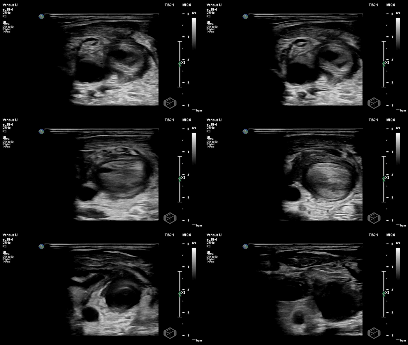
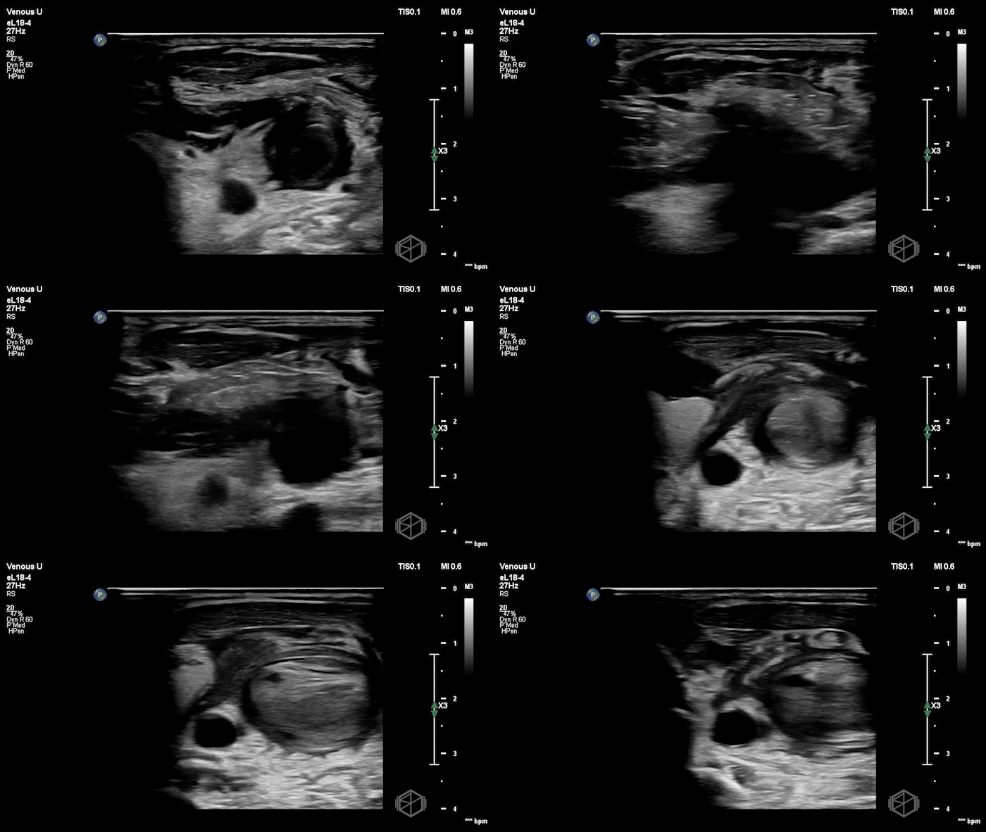
POCUS was performed within minutes of the patient’s arrival and showed the following:
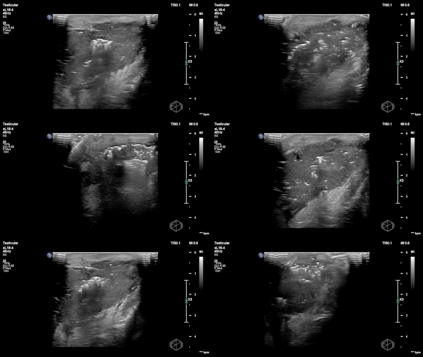
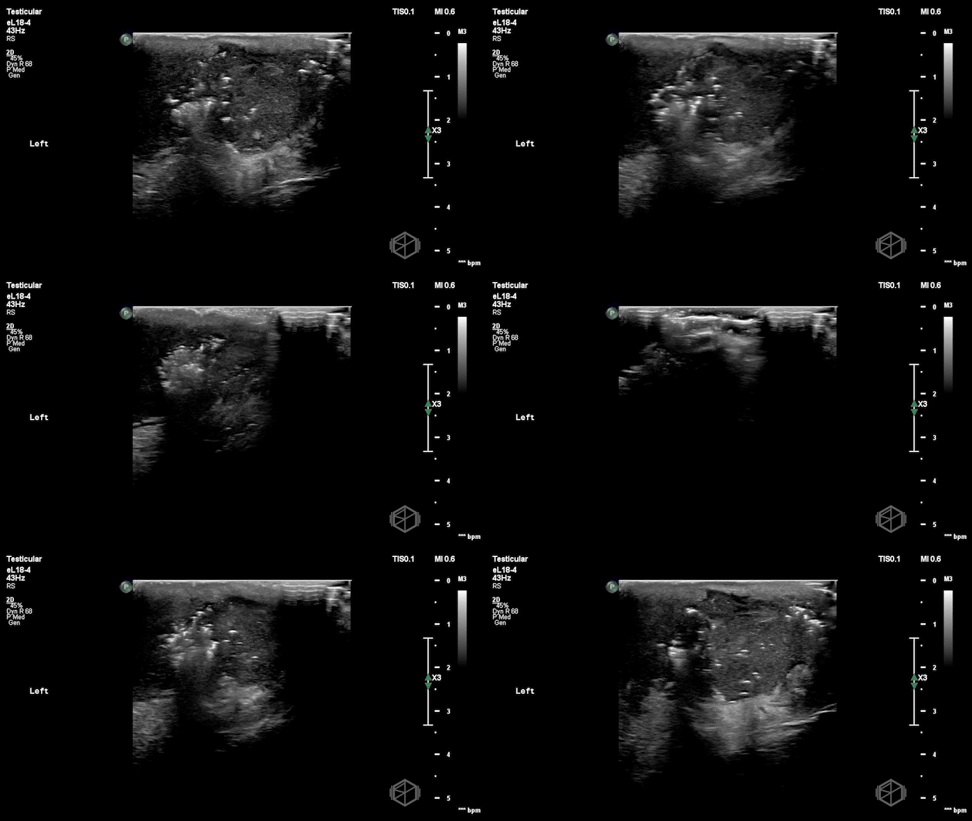
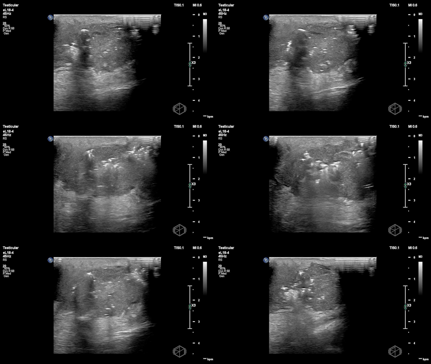
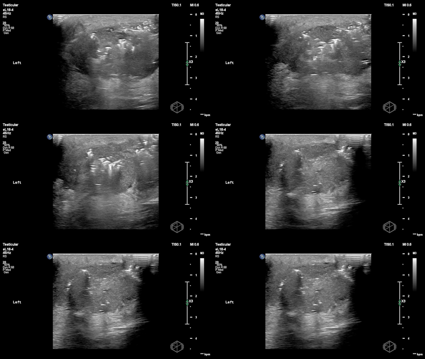
The clips show marked scrotal wall thickening and edema with extensive soft-tissue gas, seen as echogenic foci with dirty shadowing and reverberation artifact. There is also fluid tracking along the fascial planes. Findings are highly concerning for Fournier gangrene.
Blood cultures were obtained and broad-spectrum antibiotics were started. Urology was consulted and recommended further imaging.
Radiology scrotal ultrasound was read as showing bilateral hydroceles without other acute scrotal abnormality.
CT obtained later demonstrated a gas-containing collection in the left scrotum extending into the perineum, concerning for Fournier gangrene.
The patient was taken to the OR for surgical exploration. Incision of the perineum produced immediate drainage of purulent fluid with underlying necrotic tissue. Further exploration demonstrated an additional pocket extending into the inferior left hemiscrotum, requiring additional debridement.
Diagnosis: Fournier gangrene / necrotizing soft tissue infection of the scrotum and perineum
Learning Points:
Fournier gangrene may not have dramatic findings on initial exam. Pain, swelling, tenderness, or skin discoloration may precede crepitus or obvious tissue necrosis. A recent umbrella review found that up to 40% of patients may initially lack cutaneous findings, contributing to delayed diagnosis. (📚 PMID: 41439154)
Soft-tissue gas on POCUS should raise immediate concern for necrotizing infection. A small systematic review of POCUS for necrotizing fasciitis found subcutaneous emphysema to be the most specific ultrasound finding (100%; 95% CI 92.5–100%), while fascial fluid was the most sensitive (85.4%). Gas may be absent so a negative ultrasound does not exclude the diagnosis. (📚 PMID: 36580698)
The ultrasound exam should extend beyond the testes. Fournier gangrene primarily involves the scrotal and perineal soft tissues, so a testicular-focused exam may not catch the relevant pathology. Bedside ultrasound can rapidly identify soft-tissue abnormalities and help distinguish Fournier gangrene from mimics such as scrotal edema or cellulitis. In this case, radiology scrotal ultrasound showed no acute pathology beyond hydroceles, while POCUS of the symptomatic soft tissues had already demonstrated gas. (📚 PMID: 16032628)
POCUS can identify Fournier gangrene before comprehensive imaging is available. CT can help define the extent of disease and identify associated findings such as fascial thickening, inflammatory stranding, gas, fluid collections, or abscesses. With this case, POCUS demonstrated soft-tissue gas within minutes of arrival, prompting antibiotics and Urologic consultation, while CT later clarified the extent of perineal involvement. When clinical suspicion for necrotizing infection is high, particularly in an unstable patient, additional imaging should not delay surgical management. (📚 PMIDs: 35226850, 32342151, 30564282)
Time to surgical debridement matters. A systematic review and meta-analysis of 6,051 patients with necrotizing soft-tissue infection found mortality of 19% when surgery occurred within 6 hours of presentation compared with 32% when surgery was delayed beyond 6 hours. Fournier gangrene requires early surgical evaluation, broad-spectrum antibiotics, and operative source control. (📚 PMID: 31921330)
