
March SonoProps
Picking the winners of this month’s SonoProps. There were some amazing scans. Especially by Dr. Hardeep Singh, who gets the first SonoProp this month. He had a variety of great scans during his rotation with interesting pathology.
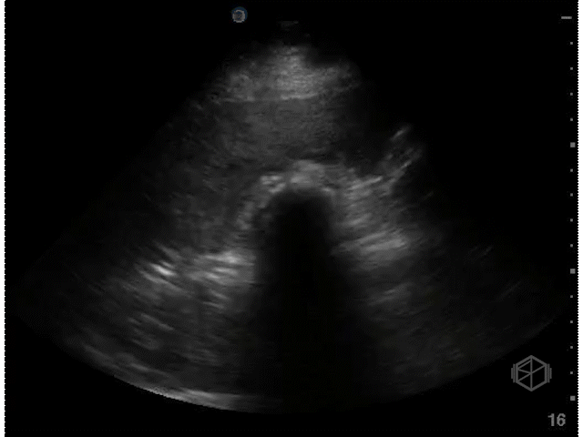
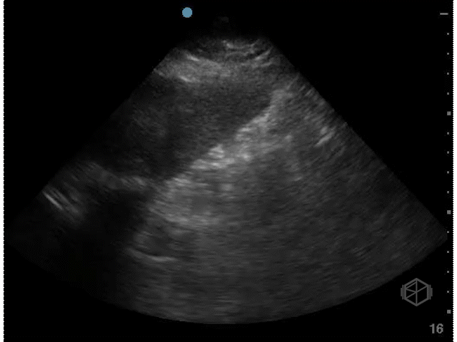
Dr. Singh scanned a ~40-year-old male with a past medical history of diabetes, hypertension, and substance abuse with diminished vision for 3 weeks.
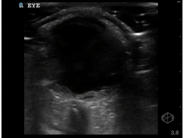
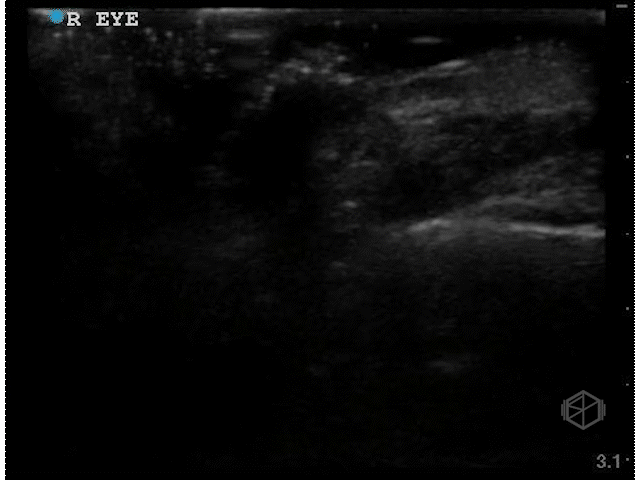
These clips are good — there is a scan both ways through the eyes in transverse and sagittal orientations. There appears to be something irregular the posterior aspect of the vitreous, however there is not enough detail.
Dr. Singh appropriately did the next step — he increased the gain significantly and performed oculo-kinetics (have the patient move their eye rapidly).
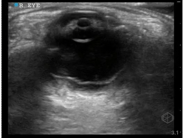
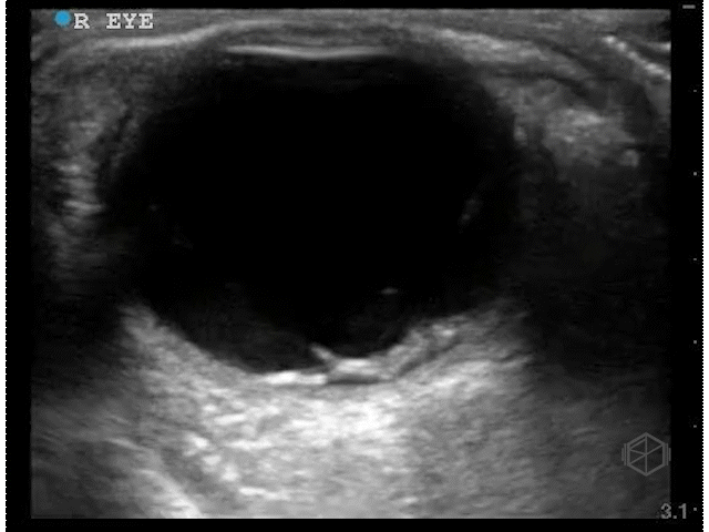

The above clips demonstrate some interesting pathology. There appears to be a hyperechoic flap tethered to the optic nerve. This patient has a retinal detachment. There may also potentially be a vitreous detachment.
Learning points:
Performing ocular ultrasound requires a few steps —
Use a high frequency linear probe. New evidence suggests that a Tegaderm is not necessary and may impede diagnostic accuracy.
Scan through the eye and saving clips both directions (transverse and sagittal).
Turn the gain up (hyper-gain) and scan again, this significantly increases the sensitivity for detection of ocular pathology.
Perform oculo-kinetics and save clips. It is important to perform oculo-kinetics as this will help determine the characteristics of any flap or pathology noted. Oculo-kinetics can help differentiate retinal detachments, vitreous detachments, choroidal detachments etc.
Optic nerve sheath diameter can be measured if necessary.
Remember to look for other pathology such as retrobulbar hematomas posterior to the eye, cellulitis, or foreign bodies when clinically indicated.
Retinal detachment appears as a thick hyperechoic membrane. It can be tethered to the optic nerve if large, or if smaller (or not close to to the optic nerve), attached to another portion of the posterior wall of the eye. The retinal detachment will move slightly with oculo-kinetics but will remained tethered. POCUS has a high sensitivity and specificity if done correctly for the detection of retinal detachments.
It is important to scan the eye in multiple planes to detect retinal detachments.
Acute retinal detachments require emergent ophthalmology consultation as there is risk for permanent vision loss and there may be ways to preserve vision. This patient likely had his retinal detachment occur 3 weeks ago.
There are more advanced aspects that can be evaluated such as mac-on and mac-off.
The next SonoProp goes to Dr. Frances Rusnack and Dr. Vivek Sharma. They scanned a ~60-year-old female with no reported past medical history who had right upper quadrant pain for 3 days and saw the following:
This is a great gallbladder scan that demonstrates acute cholecystitis. There is notable gallbladder wall thickening, pericholecystic fluid, and a hydropic gallbladder that is highlighted very well here.

CT of the same patient showing significantly distended/hydropic gallbladder with pericholecystic fluid.
Learning points:
A hydropic gallbladder (in adults) is one that measures over 10x5cm, it is one of the six signs of potential cholecystitis. There is no one sign that will ultimately definitively diagnose cholecystitis, but multiple signs together with clinical correlation help make the diagnosis.
A wider gallbladder (>5cm) is considered worse than an elongated one and portends a higher complication rate such as necrosis.
This patient’s gallbladder was longer than 11x~6.5cm, significantly hydropic. The operative note mentioned that the gallbladder was acutely edematous with pericholecystic fluid and necrosis of part of the anterior wall of the gallbladder. There were multiple large impacted stones in the gallbladder that required widening the extraction site incision.
Gallbladder hydrops usually occurs due to poor drainage of the gallbladder often caused by an obstructing stone near the cystic duct (but can occur for a number of other reasons including malignancy, cystic fibrosis, Kawasaki disease, ascariasis etc.). Bile reabsorption occurs slowly and the gallbladder wall releases mucoid or clear content which leads to distention. True hydrops occurs when the gallbladder distends without signs of inflammation.