April SonoProps
April is here, and so is another edition of SonoProps. There were a lot of great scans this month.
The first SonoProp goes to Dr. Vivek Sharma and Dr. Edward Mintz.
Dr. Mintz had a case of a 70-year-old female who presented abdominal distention and greenish non-bloody vomiting with constipation. The patient had a large palpable abdominal hernia. The patient was hypotensive on arrival and had a lactate of 2.3. Dr. Mintz performed an echo that was grossly normal so we looked for other sources. Dr. Sharma helped us perform an abdominal scan that showed the following clips.
This patient has dilated loops of small bowel, as identified by the prominent plicae circularis (“keyboard” sign), with decreased peristalsis and to-and-fro motion of bowel contents.
Within the hernia sac there is a transition point with bowel wall edema. There is also free fluid within the bowel segments suggestive of high grade obstruction (“tanga sign”).
Diagnosis: High-grade small bowel obstruction
The surgery team was called after the POCUS, before CT, and the surgical attending asked for an evaluation of blood flow which I performed.
At the time of evaluation, there was still flow present to the bowel wall. However, bowel wall edema was present which is a sign of ischemia.
The patient was admitted to the surgical service before CT and due to declining status was emergently taken to the operating room bypassing CT where she had a robotic laparoscopic small bowel resection, was left open, had a perilous course, after improving was later taken back to the operating room and had small bowel anastomosis performed. The patient tolerated diet, and was eventually discharged to home with home care.
Learning points:
POCUS for small bowel obstruction is performed using a low frequency probe (curvilinear preferred, or phased array).
Start the right lower quadrant and proceed in a lawn mower fashion up and down evaluating for dilated loops of small bowel (look for plicae circularis).
Small bowel obstruction is suggested when the following are present:
Dilated loops of bowel >2.5cm
“Keyboard” sign — well defined plicae circularis
To-and-fro motion
High-grade obstruction is suggested by (15093230):
Bowel wall edema >3mm
Absent peristalsis
Free fluid between bowel segments — “Tanga” sign
The diagnosis of a definite small bowel obstruction using ultrasound requires the identification of a transition point which is often the tricky part. Non-obstructive ileus may have some similar findings, except for a transition point, and is a potential false positive.
Keeping in mind operator dependency — POCUS is specific and accurate for the diagnosis of SBO and outperforms abdominal X-ray (29850250, 30762916, 20732861).
It can significantly reduce diagnostic time — in one study average time to CT scan report was 3 hours, 42 minutes; getting an abdominal radiograph took 1 hour, 38 minutes; and mean elapsed time to complete point-of-care ultrasonography was 11 minutes (31350094).
In another study, four 3rd year residents with 3 hours of didactics and 3 hours of practice scanning had a sensitivity of 97.7% and a specificity of 92.7% (20216422). Our residents could probably beat those numbers.
While CT cannot be replaced entirely, POCUS can expedite care, reduce disposition time, and potentially reduce unnecessary radiation.
Great job team!
Our next SonoProp goes to Dr. Yanal Maher.
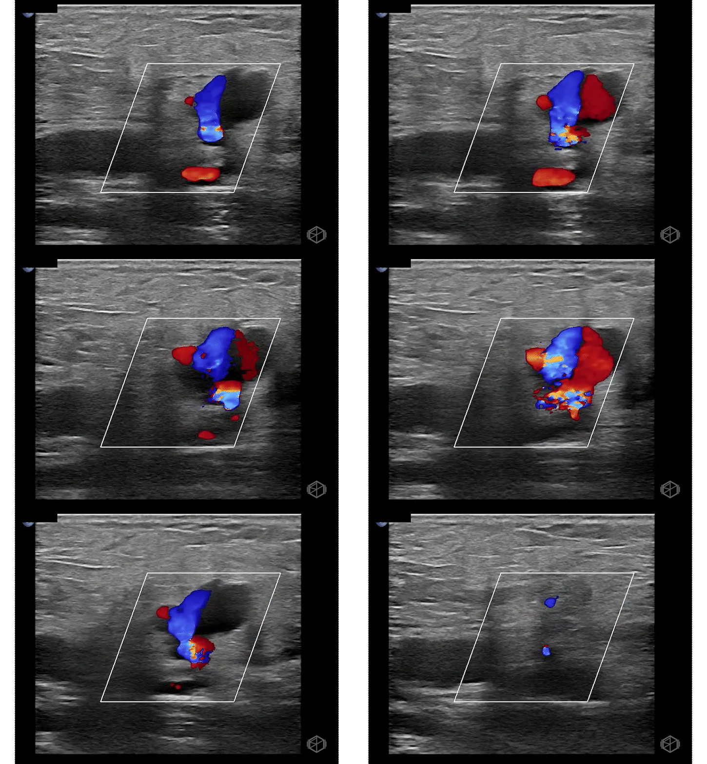
He had a 76-year-old male with a history of atrial fibrillation who recently had an ablation procedure through the right groin. The patient had an abnormal outpatient ultrasound and was referred to the ED. Dr. Maher did his own POCUS of the right groin area and saw:
Differentials for an anechoic space such as clip 1 are a hematoma, abscess, and pseudoaneurysm. The circular structure with a stalk is coming off of an artery. There is pulsatile bidirectional flow.
The findings are consistent with a femoral artery pseudoaneurysm.
Learning points:
Pseudoaneurysms, unlike true aneurysms which contain all three layers of the vessel, contain only the media or adventitia.
Pseudoaneurysms can cause significant morbidity due to risk of rupture, thromboembolism, extrinsic compression of nearby neurovascular structures, or necrosis of overlying skin and subcutaneous tissue.
Arterial pseudoaneurysms occur most often as a result of iatrogenic injury, but can also occur due to trauma, infection, or failure of an anastomoses.
Risk factors include older age, lower femoral arterial puncture, larger cannula size, and anticoagulation.
Iatrogenic causes include multiple attempts, inadequate compression post-procedure, accidental arterial dilatation during venous cannulation, and failure of closure devices.
Adequate compression post-procedure is often enough to prevent pseudoaneurysm formation.
Using the linear probe, evaluate for a pulsatile saccular structure coming off of an artery. Pseudoaneurysms may be completely anechoic or partially thrombosed. Visualize in two dimensions.
Pseudoaneurysms have a neck (or stalk, or channel) that allows blood flow into a saccular area. The neck/stalk must be visible for the diagnosis of a pseudoaneurysm (clip 3).
Due to the turbulent swirling flow, a finding known as the “yin-yang” sign may be noted. Note the “yin-yang” sign may be visible in large true aneurysms as well due to partial thrombosis.
Great job Dr. Maher!


