August SonoProps
This month was filled with good scans, but here are the SonoProps:
The first SonoProps award goes to Dr. Andrew Weinberger.
Dr. Weinberger evaluated a woman in her 60s with a complex past medical history, including a recent GI bleed treated with open small bowel resection for a bleeding AVM. Prior to surgery, multiple endoscopies had failed to identify the source of bleeding. A nuclear medicine bleeding scan eventually localized it to a distal jejunal loop, prompting surgery. The patient recovered well postoperatively and was observed for a period before discharge.
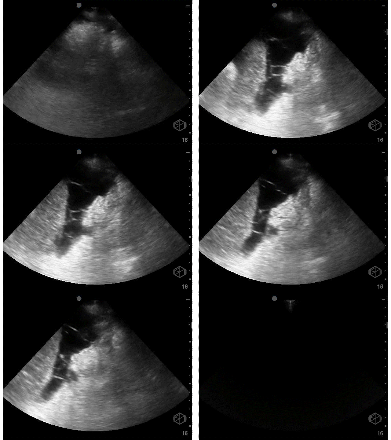
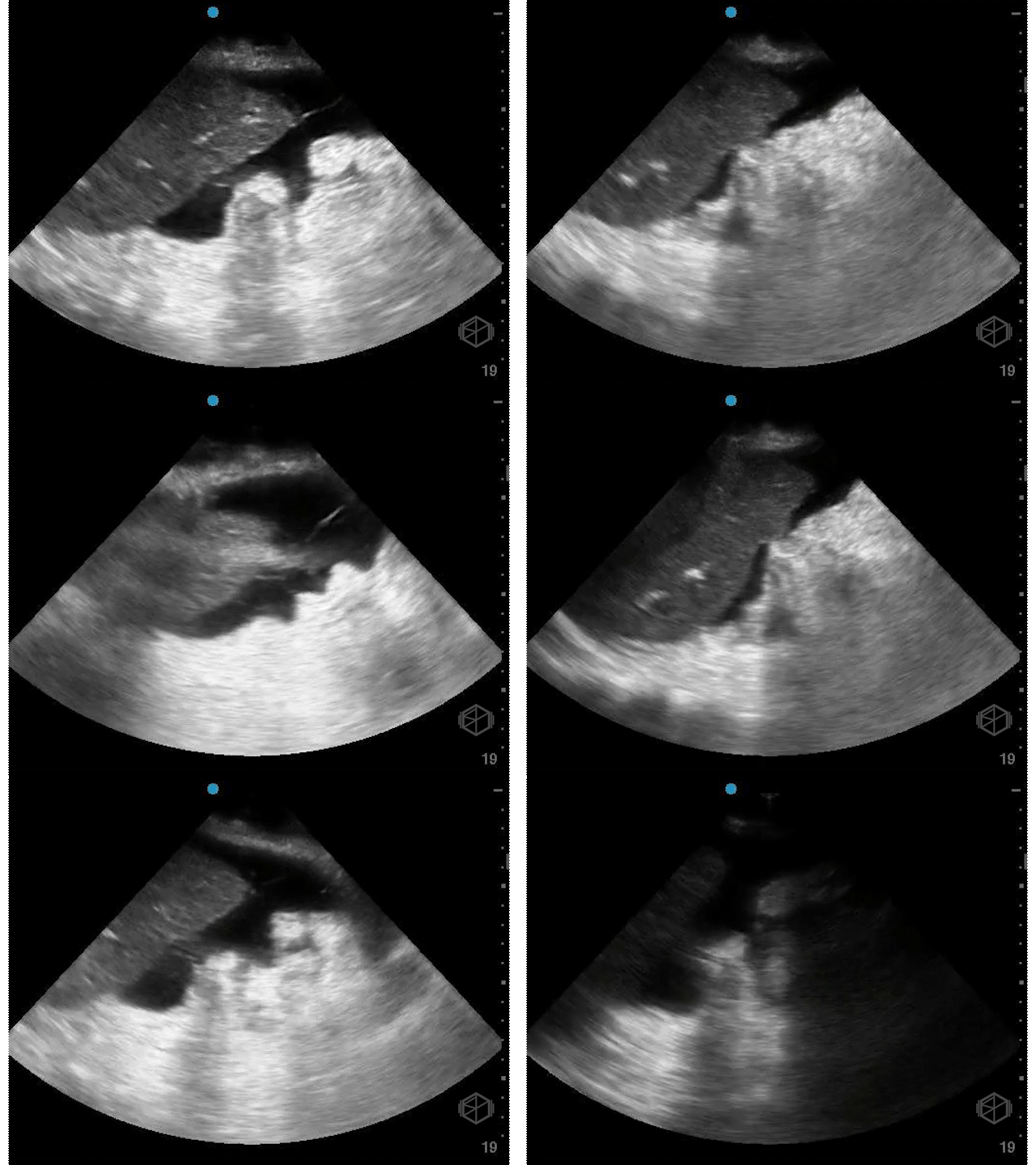
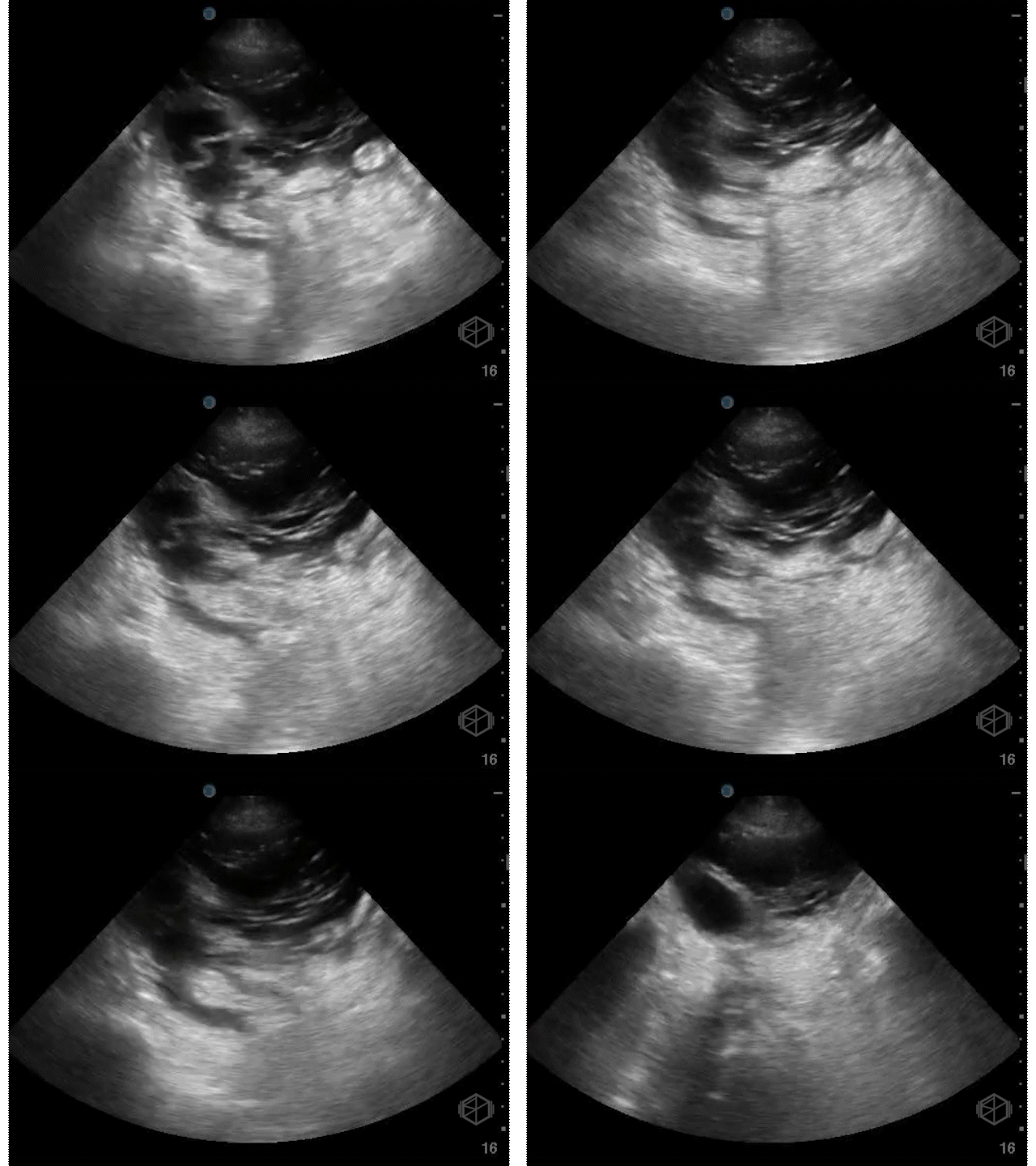
Several days later, she returned to the ED with hypotension, lightheadedness, and mild abdominal discomfort. Recognizing the potential urgency, Dr. Weinberger immediately performed a POCUS, which revealed the following:
The patient had complex free fluid in the abdomen, noted with septations and fibrinous stranding. Surgery was immediately notified and a CT was performed a little while later that showed a moderate amount of free air in the abdomen, fluid suspicious for peritonitis, and concern for a leak at the small bowel connection in the right lower quadrant. In surgery, cloudy bile-tinged fluid was found but no stool. The previous connection had poor blood flow and a small leak. That section was removed, a new connection was made between healthy bowel, and the abdomen was closed. The patient tolerated the procedure and did well.
Diagnosis: Complex free fluid due to small bowel anastomotic leak
Learning points:
In this case, bedside ultrasound quickly detected complex free fluid, which elevated the urgency and directly led to prompt CT and surgical involvement—without waiting for lab results or other imaging. This aligns with literature establishing POCUS as an important diagnostic procedure that goes beyond clinical examination and streamlines critical decision-making (📚 PMID: 30356808).
A good editorial written by surgeons, emphasized the value of POCUS for detecting intra-abdominal free fluid and expediting decision-making in acute abdominal cases, noting its role in “ruling in” pathology when performed by skilled operators (📚 Signa Vitae, 2023).
On POCUS, complex free fluid often appears echogenic, may contain septations, debris, or a layered appearance—much different from simple anechoic fluid. This sonographic appearance commonly indicates infection, blood, or bile, and in unstable postoperative patients, any complex fluid should be considered pathologic until proven otherwise. (📚 PMID: 28567101)
While fluid echogenicity alone doesn’t always distinguish cause, clinical context and POCUS features help guide rapid triage (📚PMID: 39640082).
Early diagnosis and timely surgical intervention in postoperative peritonitis are firmly linked to improved outcomes. Catching these early and is very important, and POCUS can play a part in that. Delays in relaparotomy are associated with significantly higher morbidity and mortality (📚PMID: 19948445).
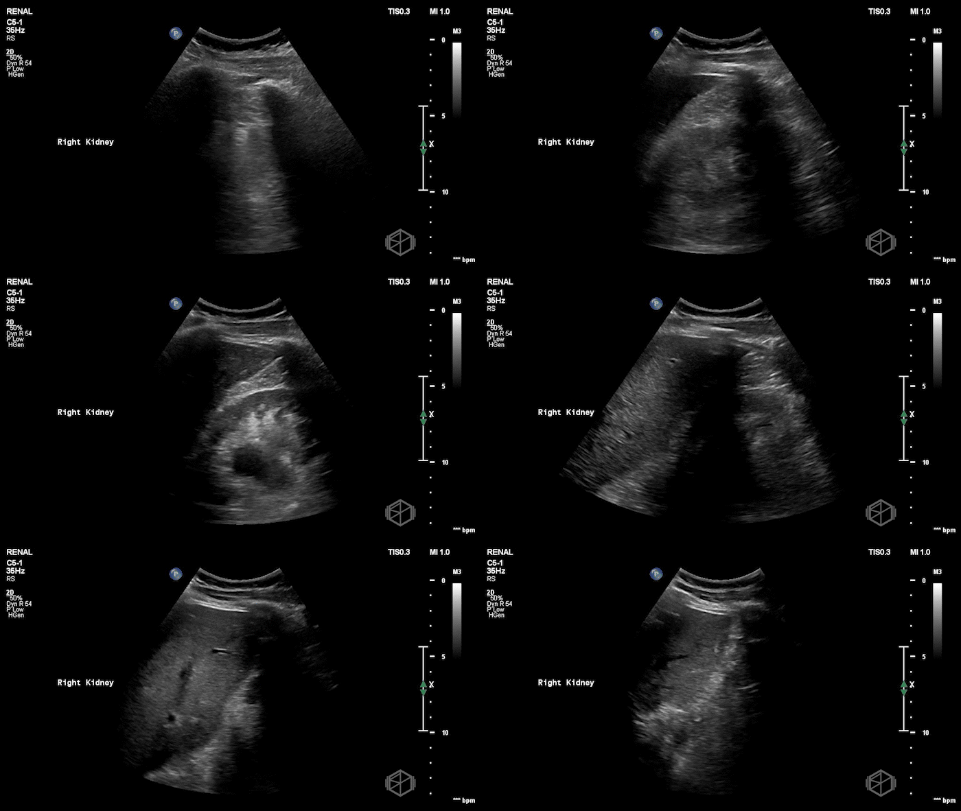
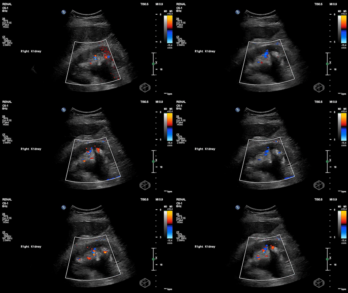
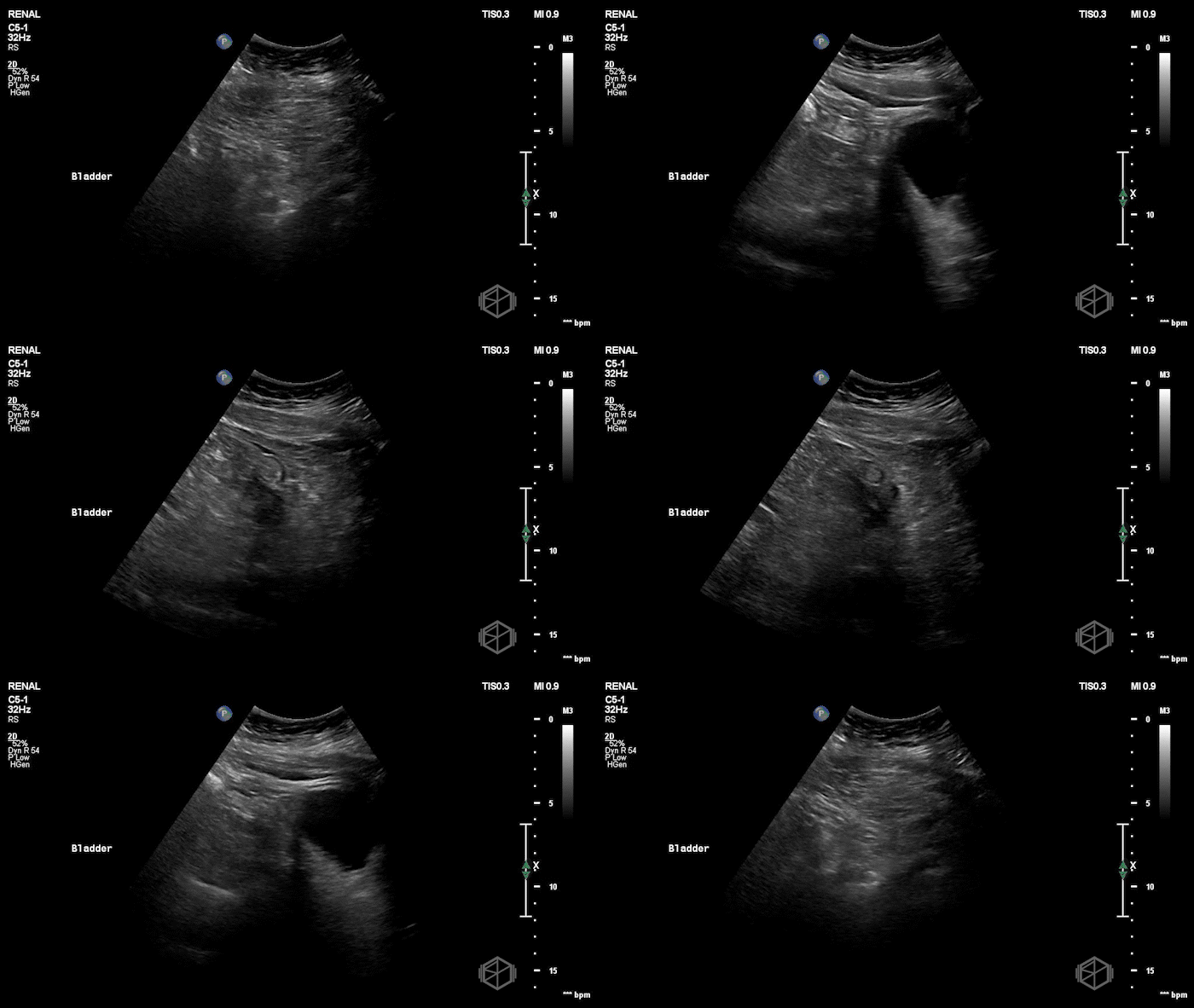
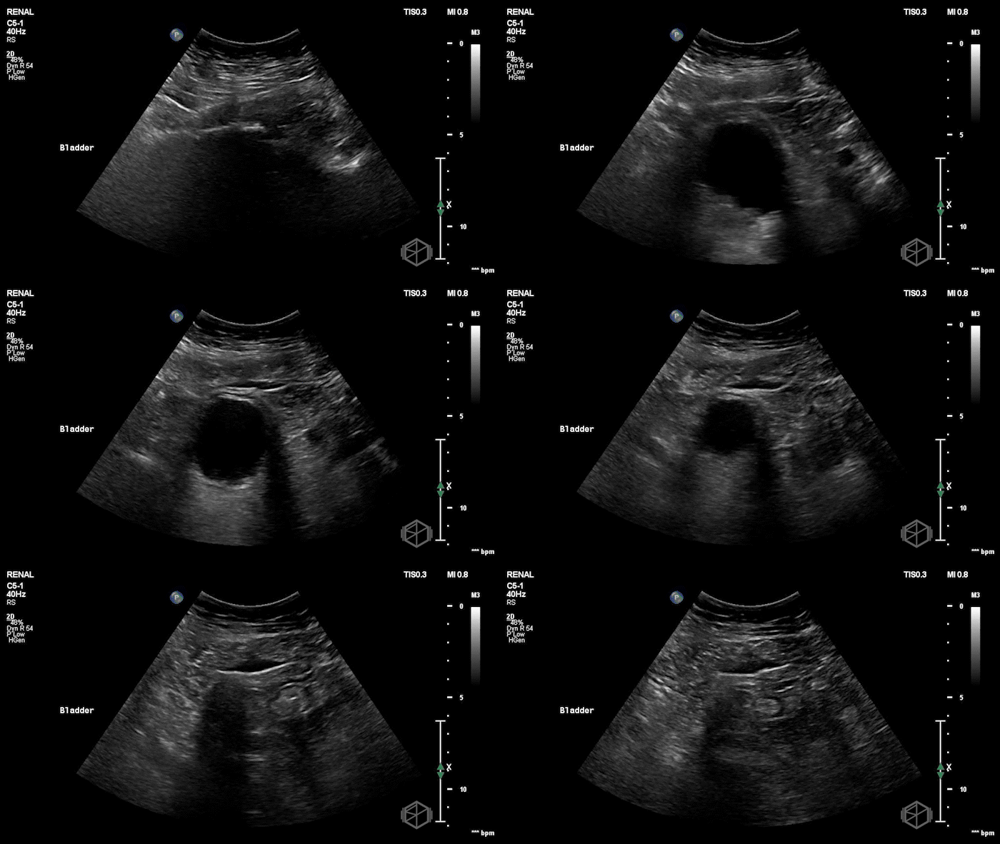
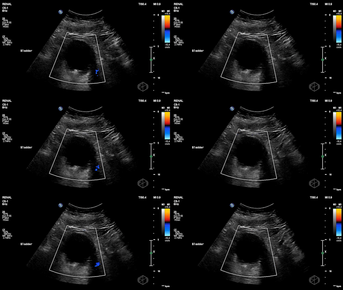
A 70's male with a history of nephrolithiasis presented to the ED with right flank pain worse over the last 4 days. Dr. Mendelow performed a POCUS immediately that showed:
Subsequent CT scanning later showed a 10 x 7 x 7mm right ureterovesical junction calculus within the urinary bladder causing moderate right hydroureteronephrosis, delayed nephrogram and mild perinephric fat stranding. The patient was taken to the OR for scope of bladder and right ureter; laser lithotripsy and removal of right ureteral stone and placement of right ureteral stent.
Diagnosis: 10mm UVJ stone with moderate hydronephrosis
Learning points:
Safe “US-first” pathway doesn’t miss bad outcomes. In a multicenter RCT (n=2,759), an ultrasound-first strategy cut radiation exposure without increasing missed high-risk diagnoses, serious adverse events, ED revisits, or hospitalizations vs CT-first (📚 PMID: 25229916)
Not all stones twinkle — Twinkle artifact has limitations. Doppler twinkling artifact has low sensitivity (~54%) but high specificity (~95%) for detecting urinary stones, meaning a missing twinkle does not rule a stone out
(📚 PMID: 31700749).
Shadowing and hyperechoic focus matter. Gray‑scale ultrasound findings such as bright (hyperechoic) foci with posterior acoustic shadowing often precede suspicion of stones—especially larger ones or those at the ureterovesical junction.
Color Doppler absence of ureteral jets may suggest obstruction. Most healthy people have a ureteric jet frequency of 2 or more per minute on either side. One study reports sensitivity ~87%, specificity ~96% for detecting ureteral obstruction when less than 25% ejections are seen over five minutes, another found sensitivity to be 94.8% but specificity to be 55.5% (📚 PMID: 37465268).
The practical utility is debated—bilateral strong jets are reassuring, but absence is only meaningful when there’s a high suspicion for unilateral obstruction and no jet is seen after adequate observation. There is no need to wait any longer than 2 minutes at the most (📚 PMID: 16509793).
Jets may be absent when renal plasma flow is reduced or when one kidney’s blood supply is impaired (eg, in renal cell carcinoma with renal vein blockage from tumor invasion or clot).


