September SonoProps
September is here and so is a new duo of SonoProps!
Our first SonoProps goes to Dr. Yianni Flouskakos.
Dr. Flouskakos is on the US rotation currently and was scanning a late 80’s female with mild left lower quadrant and left flank pain when he noticed the following findings:
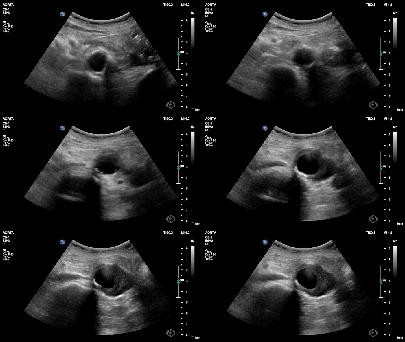
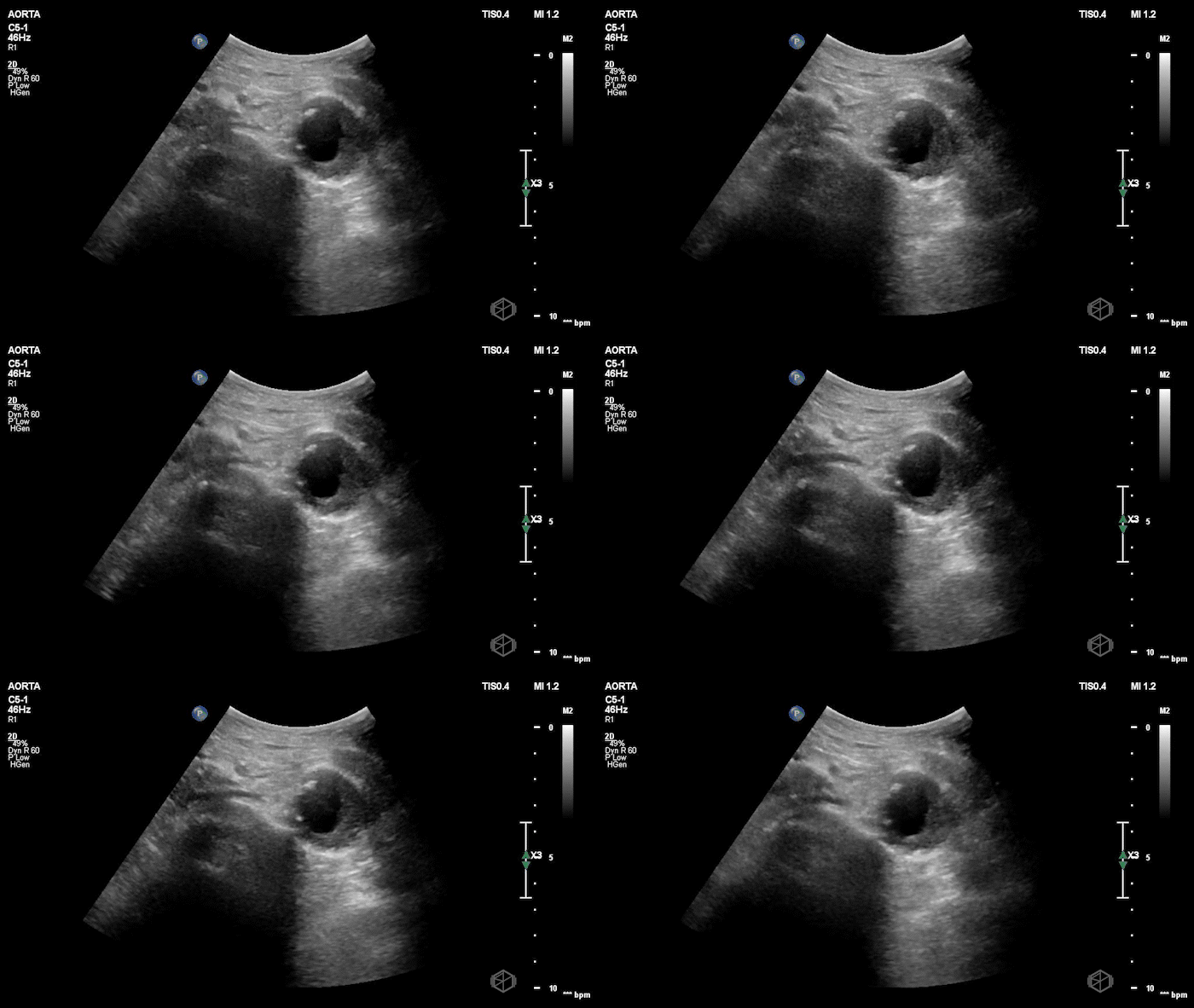


Upon scanning the aorta, it was noted that the patient an enlarged infra-renal abdominal aorta with an eccentric left thrombus, measuring approximately 4.05cm consistent with an abdominal aortic aneurysm. On lateral view the aneurysm appears more saccular than fusiform. CT was done which confirmed the finding: Saccular infrarenal abdominal aortic aneurysm with partially thrombosed aneurysm sac eccentric to the left measuring up to 4.0 cm.
Diagnosis: Saccular aortic aneurysm
Learning points:
POCUS can be valuable in symptomatic elderly patients with abdominal, flank, or back pain, where differentials include diverticulitis, renal colic, or AAA. A quick bedside look at the aorta can dramatically shift management. (📚PMID: 23406071)
POCUS is highly accurate for detecting AAA in the ED. A 2024 meta-analysis (9 studies) found pooled sensitivity 98.3% and specificity 99.8% for ED POCUS diagnosing AAA (📚PMID: 39344280)
POCUS can shorten time to diagnosis in ruptured AAA pathways. ED cohort showed a trend toward faster diagnosis with ED ultrasound (~51 min vs ~111 min). (📚PMID: 23995666)
Saccular AAAs tend to rupture at smaller diameters than fusiform. Contemporary multicenter data show ruptured saccular AAAs present at smaller sizes, supporting lower size thresholds for repair. (📚PMID: 38780177)
Women rupture at smaller sizes and have higher rupture risk. Reviews/observational data highlight sex-specific risk—supports closer surveillance and earlier consideration of repair in women. (📚PMID: 23384493)
POCUS is feasible even with limited prior experience (with training). Early ED work showed accurate scans by relative novices—useful for teaching and justifying aortic sweeps in undifferentiated pain. (📚PMID: 10969223)
The next SonoProps goes to Dr. Eytan Mendelow!
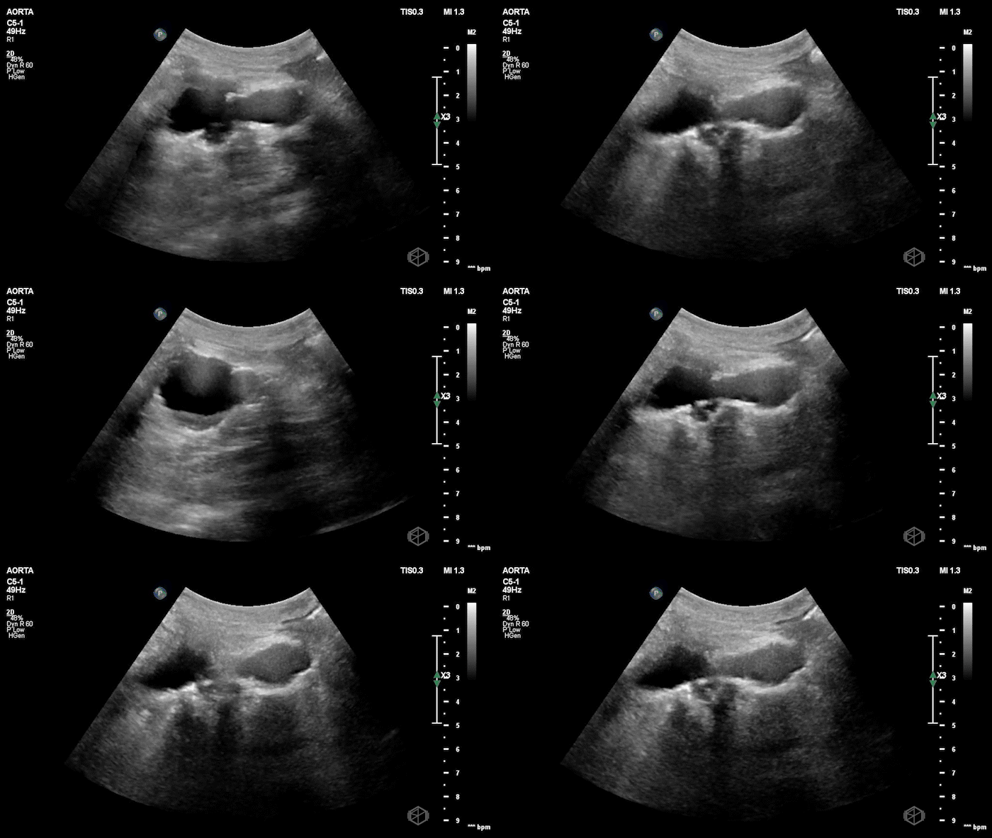
Dr. Mendelow was scanning a late 20’s male with acute epigastric and right upper quadrant pain with nausea and vomiting. He was significantly hypertensive and tachycardic likely due to pain. On exam the patient had a notable Murphy’s sign.
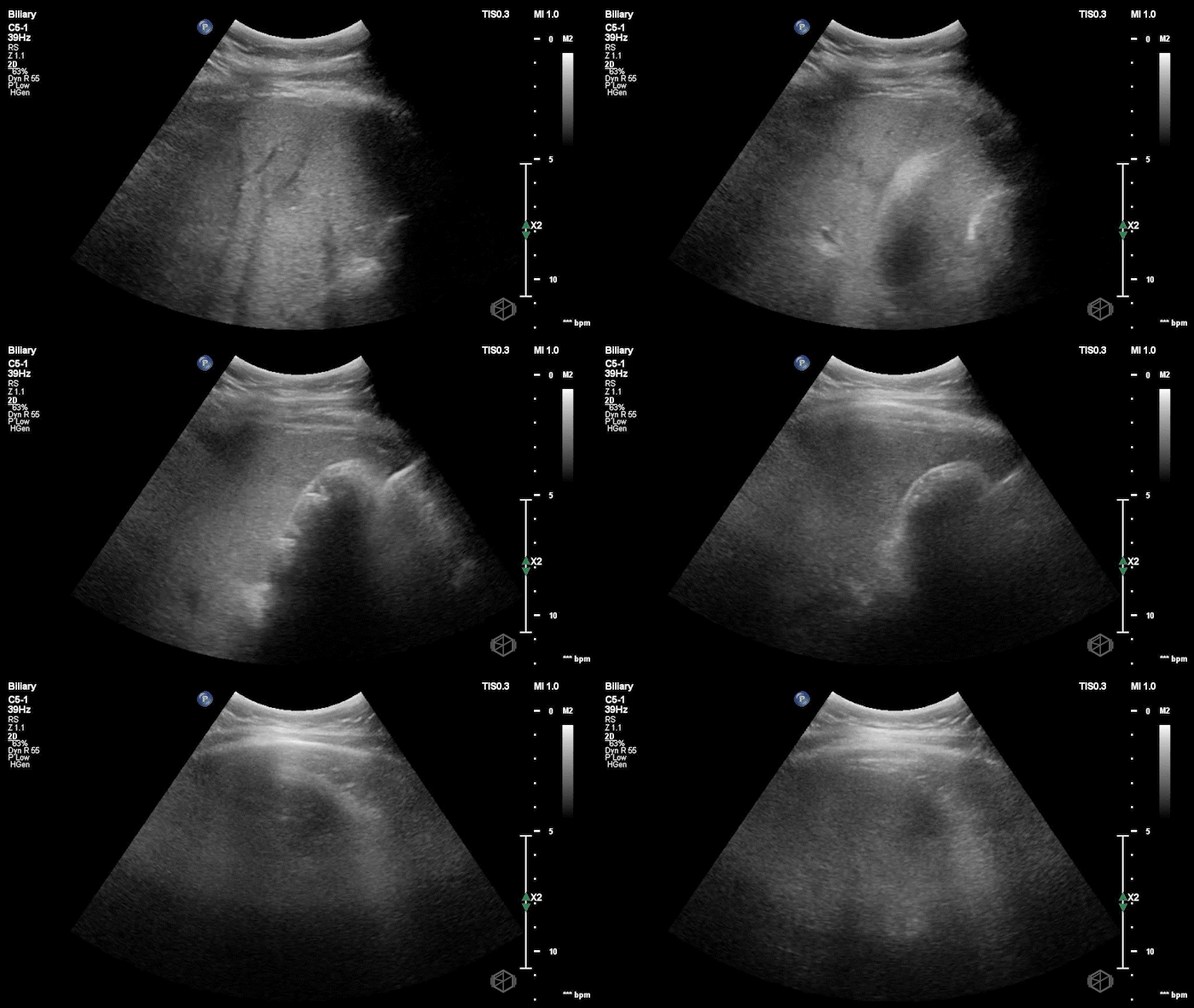
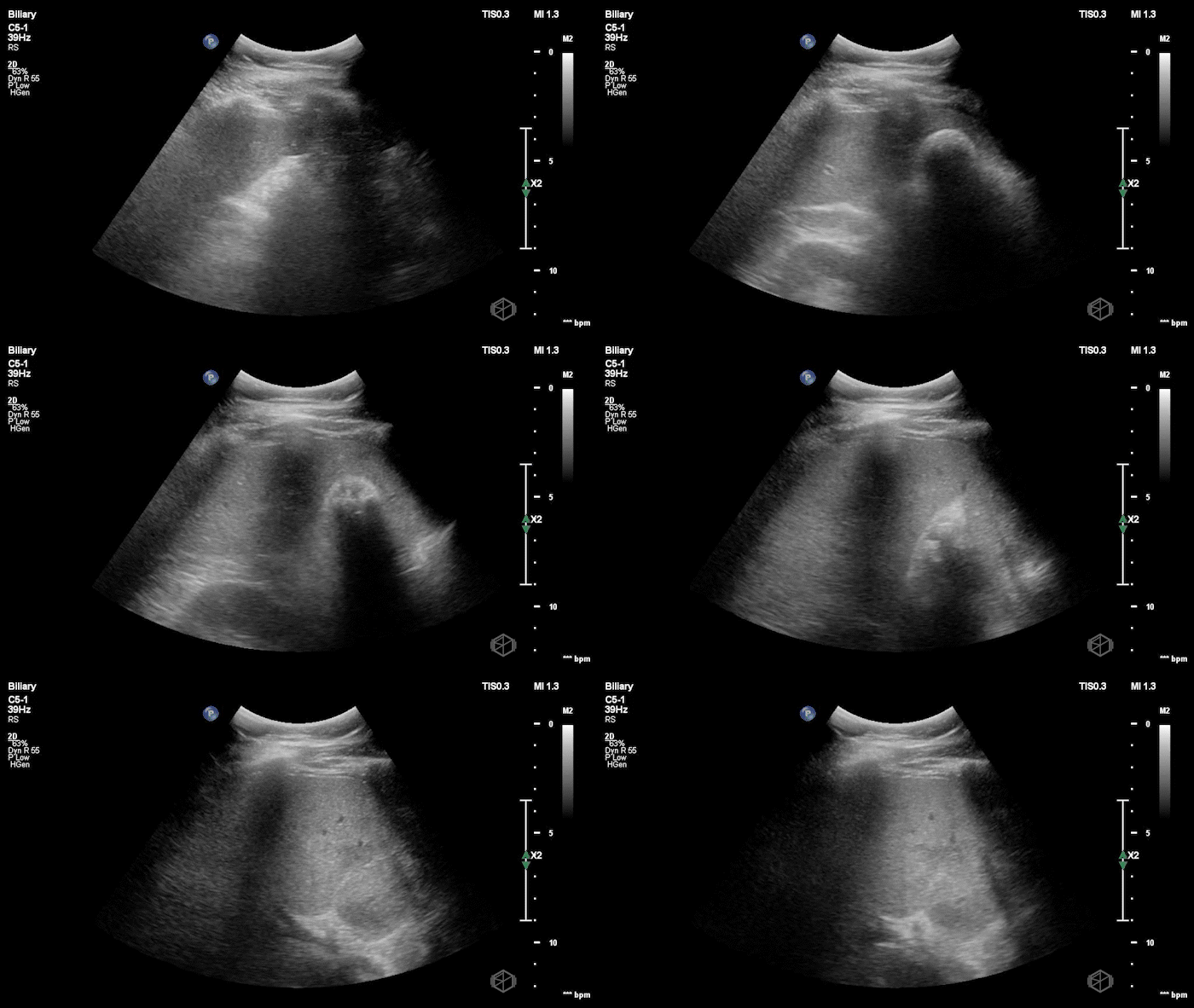
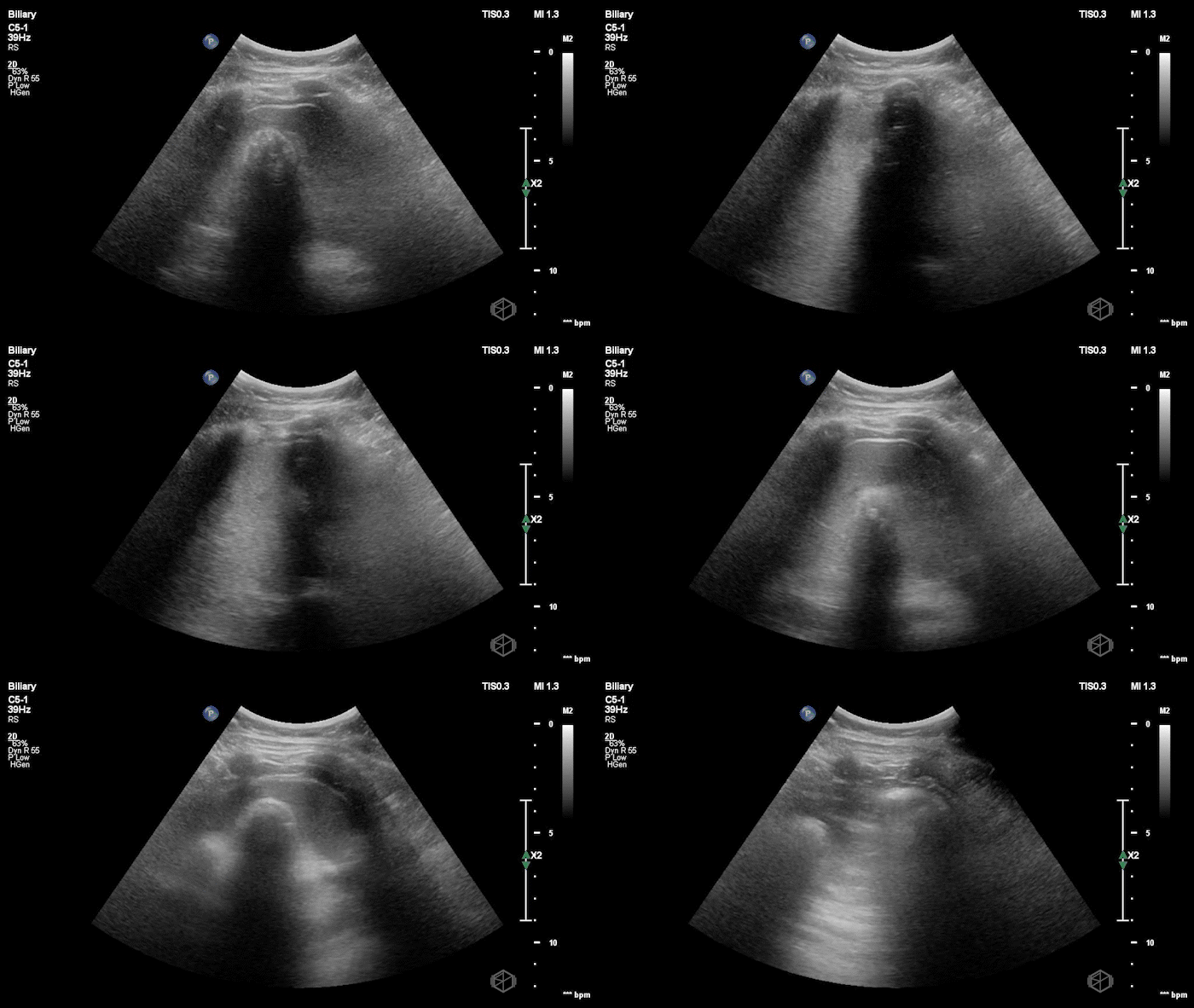
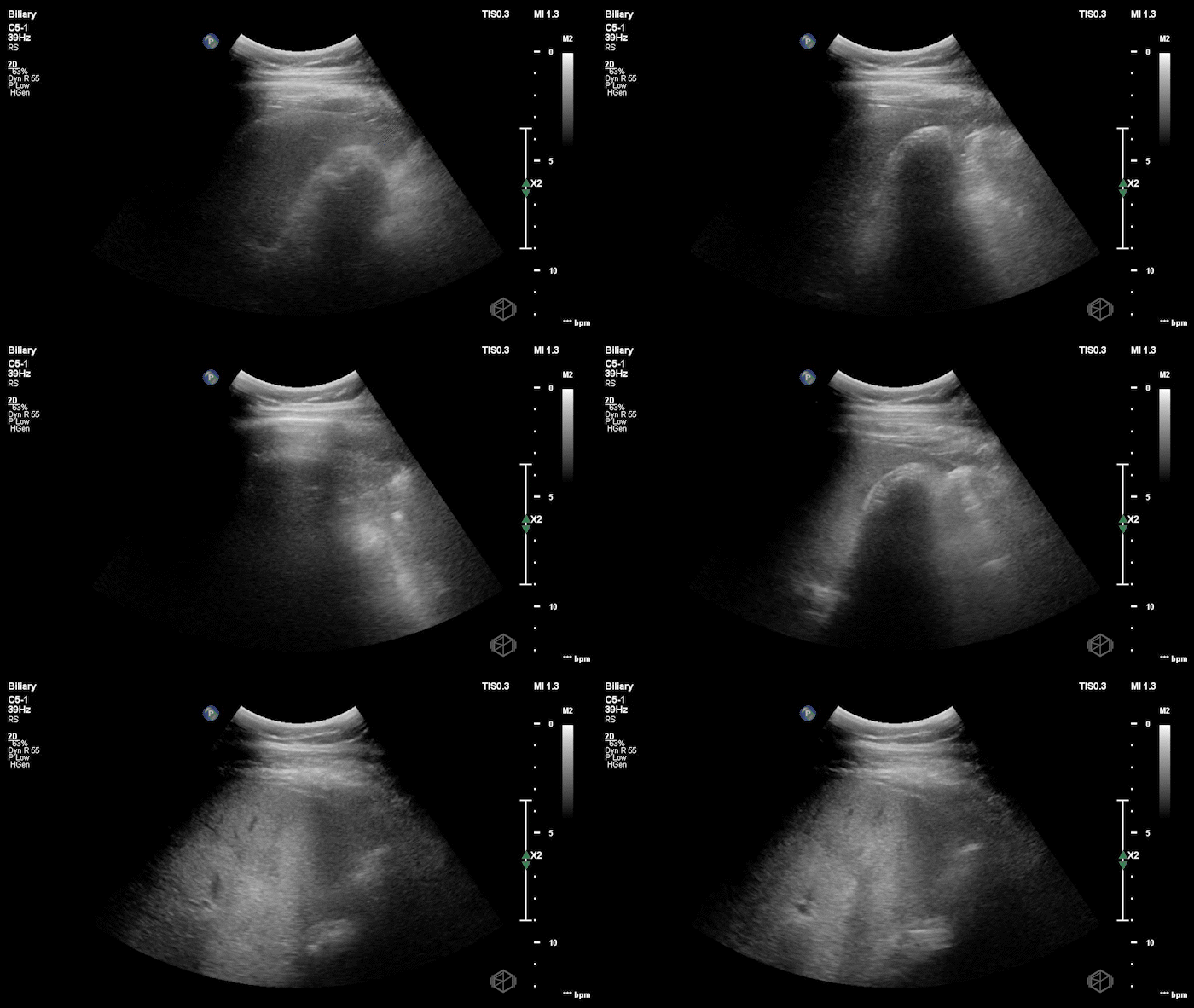
Dr. Mendelow performed a POCUS within 5 minutes that showed the following:
This scan shows a gallbladder filled with stones as indicated by the bright hyperechoic stones with shadowing posteriorly and almost no visible gallbladder wall lumen. This is consistent with a Wall-Echo-Shadow (WES) sign or configuration. The patient had a significantly elevated lipase and total bilirubin indicating gallstone pancreatitis. Radiology ultrasound resulted 90 minutes later with similar findings.
Diagnosis: Gallstone pancreatitis; WES sign
Learning Points:
WES sign: A gallbladder with three layers: bright anterior wall, immediately adjacent hyperechoic stones, and a clean posterior acoustic shadow. Lumen nearly obliterated. Classic sign of a gallbladder packed with calculi.
WES can sometimes be mixed up with porcelain gallbladder which can be tricky to differentiate. Visualization of the gallbladder wall is essential in differentiating between a positive wall-echo-shadow (WES) sign and a porcelain gallbladder. While a visible gallbladder wall is indicative of a WES sign, a hyperechoic wall layer without any obvious stones will indicate a calcified gallbladder wall, suggesting a porcelain gallbladder. (📚 PMID: 37465707)
WES ≠ cholecystitis (by itself): WES alone doesn’t diagnose acute cholecystitis. It may indicate subclinical cholecystitis, acute or chronic especially when combined with sonographic Murphy’s sign.
POCUS can stand on its own. Zitek et al. (2023) showed ED biliary POCUS is 97% sensitive and 99.5% specific for stones. When scans are positive and symptoms fit, confirmatory imaging rarely changes management. Save extra imaging for negative or equivocal cases. Ensure you view the neck of the gallbladder as that is a potential pitfall for missing stones. (📚 PMID: 36279080)
Cannata et al. (2024): More imaging = more time, less clarity. Median time to POCUS was ~115 min vs ~313 min for radiology imaging. When POCUS was positive (performed by POCUS credentialed clinicians), radiology ultrasound agreed 82% of the time, and in discrepant cases, POCUS was correct in 8/9 with surgical confirmation. Once you’ve got a solid POCUS, extra imaging often slows things down and muddies the waters. (📚 PMID: 38681169)
Huang et al. (2023): Trust the positive. Meta-analysis found ultrasound has high specificity (~90%) but only moderate sensitivity for acute cholecystitis. In practice: a positive scan with stones, Murphy, and wall changes is almost always right. (📚 PMID: 38037062)

