March SonoProps
Hello everyone, welcome to another edition of SonoProps.
This month’s first SonoProps goes to Dr. Stephen Simon.
Dr. Simon was taking care of a 60’s female patient who had a mechanical fall while carrying bags. The patient had a laceration over her left eyebrow and significant periorbital swelling and some proptosis. The cornea was cloudy and he was unable to visualize the pupil. She had pain with movement of her eye.
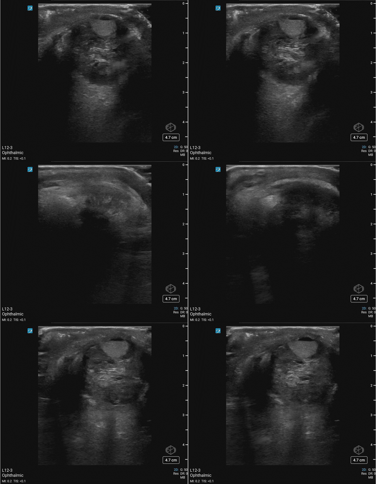
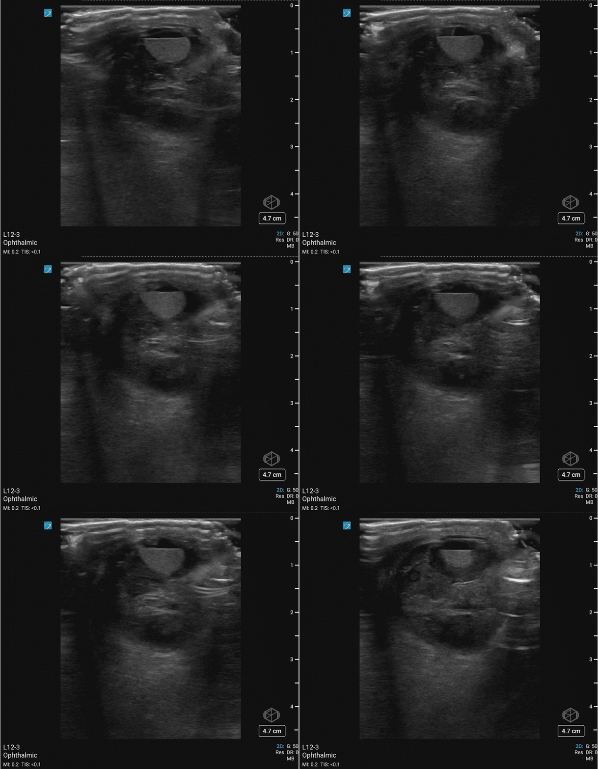
Dr. Simon very carefully performed an ocular ultrasound:
Upon seeing this he immediately terminated the ultrasound having made the diagnosis. With multiple differentials to consider this ultrasound has some great findings. The anterior chamber of the eye demonstrates layering fluid. The vitreous body is completely heterogenous instead of anechoic. There is no guitar pick sign or anechoic material posterior the eye that would indicate a retrobulbar hematoma. The patient was evaluated by ophthalmology that confirmed his diagnosis and the patient was transferred for ophthalmologic surgery.
Diagnosis: Traumatic globe rupture with significant vitreous hemorrhage and a hyphema
Learning points:
Ocular ultrasound can be incredibly valuable for traumatic injuries especially in the case of significant periorbital edema where the eye cannot directly be examined revealing potential diagnoses such as hyphema, globe rupture*, retrobulbar hematoma, and potentially foreign bodies. CT is extremely useful but POCUS can be performed at bedside.
A hyphema appears as a settled echogenic blood within the aqueous of the anterior chamber.
This patient has a significant vitreous hemorrhage/intraocular hematoma as noted by the entire vitreous body filled with echogenic material.
The globe rupture is suggested by the decreased size of the globe, indicating loss of pressure or vitreous.
If physical exam suggested an open globe, POCUS should be avoided to avoid causing further extrusion of intraocular contents. CT has a sensitivity of 75% and a specificity of 93% based on one paper (11013196). Thin-slice (2mm) increases the specificity to 97% while maintaining a similar sensitivity (28662312). If there is a high suspicion despite negative imaging, ophthalmology should be consulted.
If there is no definite evidence of globe rupture on exam then POCUS can be performed maintaining minimal pressure on the eye and should be immediately terminated (as Dr. Simon did) when open globe is suspected on POCUS. A study on health volunteers demonstrated only a minimal increase in intraocular pressure when POCUS was performed, however caution should still be maintained in trauma patients (25834668).
The second SonoProps of this month goes to Dr. Michael Gorynski.
Dr. Gorynski had a 60’s male present to the ED with mild pain and a palpable lump to the right upper extremity for the past few days prior to arrival. The exam revealed no erythema, warmth, tenderness, crepitus, deformity, or ecchymosis but there was a palpable cord next to the medial epicondyle.
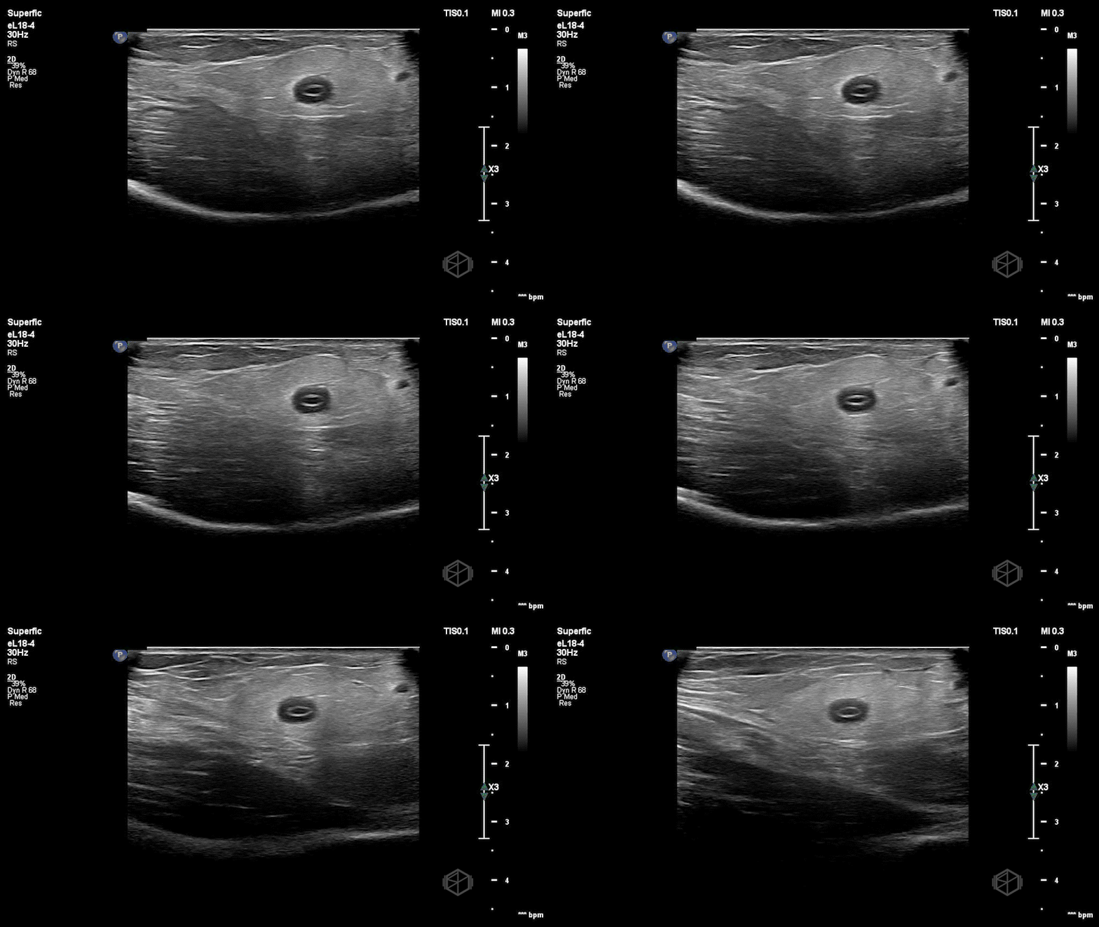
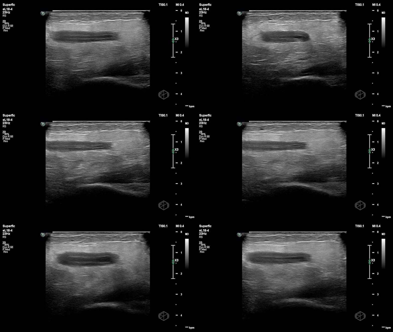
He performed a POCUS of the area that showed:
This patient has a basilic vein that has significant mural thickening with some echogenic material within the vessel in clip 2. It remains compressible. The patient was discharged with appropriate instructions. He later returned and was seen again by Dr. Gorynski a few days later for a similar complaint and had a radiology ultrasound (RADUS) that demonstrated chronic superficial thrombophlebitis involving the cephalic and basilic veins of the right upper extremity, but this visit took far longer than his first due to the RADUS.
Diagnosis: Superficial thrombophlebitis of upper extremity vein
Learning points:
POCUS for soft tissue complaints can significantly improve accuracy and rapidity of diagnosis. In this case the thrombophlebitis was diagnosed quickly by Dr. Gorynski leading to a faster disposition time.
Remember, deep veins have a corresponding named artery. In the upper extremity the deep veins include the subclavian vein, axillary vein, paired brachial veins, paired ulnar veins, and paired radial veins. The basilic and cephalic are superficial veins.
Superficial thrombophlebitis can mimic cellulitis and/or abscess but can be distinguished using POCUS.
Normal veins demonstrate an anechoic lumen with a smooth wall and complete compressibility. On POCUS inflammation/edema of the vein can indicate phlebitis and presence of echogenic material within the vessel indicates thrombophlebitis (30171620).
This patient did not have the classic erythema but did have the palpable cord on exam.
Superficial thrombophlebitis is usually self-limited and treated with warm compresses and NSAIDs (unless the patient has factors that limit the use such as renal disease).


