
November SonoProps
Hello everyone,
This month’s first SonoProps goes to Dr. Antonia Nevias-Ida, working with Dr. Zimmerman!
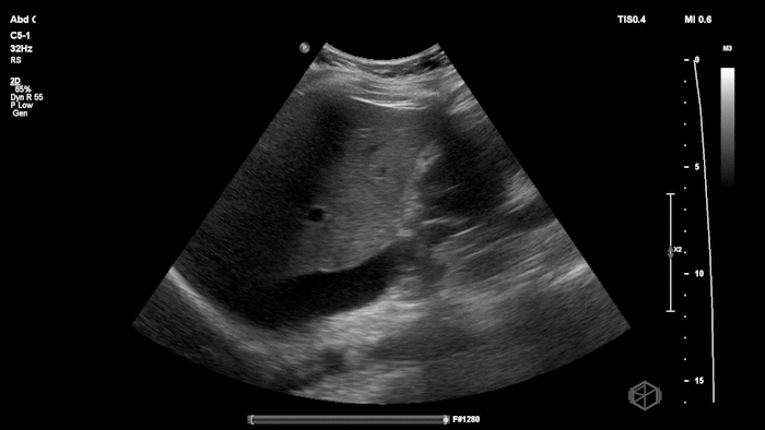
Dr. Nevias-Ida had a patient with 20’s female with PCOS who came in status post egg retrieval for IVF. The patient was complaining of abdominal pain, fullness, and nausea. She had some abdominal tenderness and distension.
She grabbed the probe, and working with Dr. Zimmerman, saw the following:
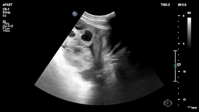

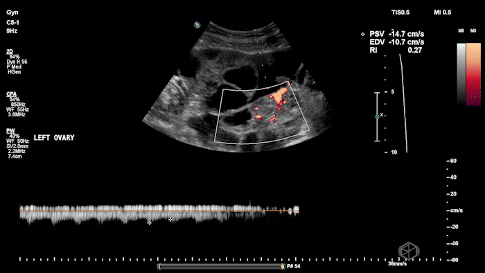
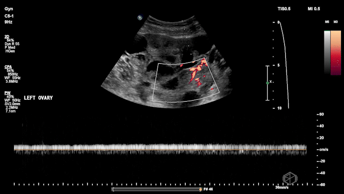
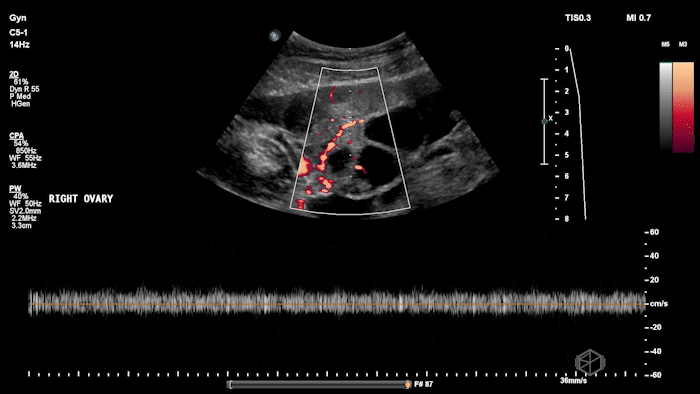
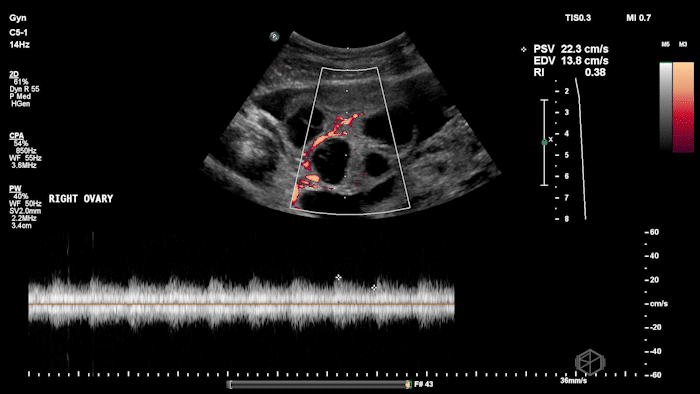
There is moderate free fluid, no obvious pleural effusions. The bilateral ovaries are enlarged with multiple cystic structures. There are normal arterial and venous waveforms for both ovaries.
Diagnosis: Ovarian Hyperstimulation Syndrome
Learning Points:
OHSS is a known complication of ovulation induction/assisted reproductive technologies. Risk factors include: PCOS, high follicle/oocyte yield, rapid ovarian enlargement. (📚 PMID: 25302750)
OHSS is possibly due to capillary leak driven primarily by VEGF, which is stimulated by hCG → fluid shifts into the third space (ascites, distension, hemoconcentration). (📚 PMID: 29892142)
RAS activation (also triggered by hCG) works alongside VEGF to worsen vascular permeability, and there are many other factors (eg, Interleukin 6, angiotensin II, and insulin-like growth factor-1).
Bottom line: hCG → VEGF + RAS → massive third-spacing.
Typical ultrasound findings: enlarged ovaries with multiple cysts/follicles; free fluid (ascites) and in more severe cases pleural or pericardial effusions. (📚 PMID: 27998636)
Preserved ovarian arterial and venous flow helps distinguish OHSS from acute ovarian torsion, where flow may be compromised.
Management varies by severity and there many classification schemes for grading the severity. (📚 PMID: 27998636)
Mild OHSS — Abdominal discomfort/bloating with little to no ascites → typically outpatient management with close follow-up.
Moderate OHSS — Increasing pain, nausea/vomiting, ascites present but stable vitals → consider observation and supportive care.
Severe OHSS — Large-volume ascites, ± pleural/pericardial effusions, hemoconcentration, tachycardia, electrolyte abnormalities → hospitalization/ICU, fluid balance monitoring, VTE prophylaxis, and paracentesis if symptomatic.
Prompt recognition and monitoring can prevent complications and guide appropriate management, great scan Dr. Nevias-Ida!
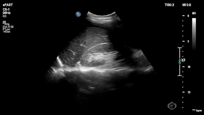
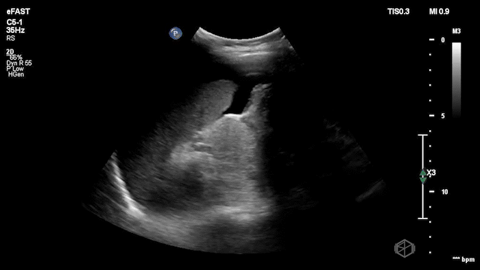
The next SonoProps goes to our current month’s rotators Dr. Marlene Konner and Dr. Yanal Maher. They were scanning a 30’s female who presented to the ED for low back pain and nausea/vomiting. On exam she had right costovertebral angle tenderness and mild diffuse abdominal tenderness.
Dr.’s Konner and Maher scanned the patient and saw the following:
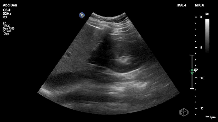
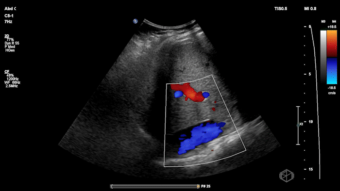
Initially it appears as though there is anechoic material above the liver and it does seem to make sharp angles. However, Dr. Konner and Dr. Maher utilized color doppler to evaluate the area and noted that it was simply the inferior vena cava. The patient ultimately had a CT scan that demonstrated no emergent findings.
Diagnosis: Potential false positive FAST - Inferior Vena Cava as a mimic of Free Fluid.
Learning points:
Vessels can mimic free fluid. The IVC and other vascular structures sit right where we look for RUQ fluid and may appear anechoic in a single still frame. Use color doppler when not sure, free fluid will most commonly be at the caudal liver tip, and rarely just above the spine where the IVC sits.
Other mimickers of free fluid in the right upper quadrant include:
Always identify known landmarks (diaphragm, liver edge, kidney, spine) in RUQ views. If you see an anechoic region, ask: Does this structure have sharp, well-defined walls (suggesting a vessel)? Does it run longitudinally/continuously (vessel) or is it pooling between structures (free fluid)?
Remember that a negative POCUS does not rule out injury: eFAST/FAST is best as a rule-in test for free fluid, not a definitive rule-out.
Great scan that will definitely be used in future presentations Dr. Konner and Dr. Maher!