General notes:
Must be in abdominal or renal setting.
Must include both kidneys (if patient has both) in two views (longitudinal and short). Measure kidney length.
Evaluate for hydronephrosis and grade. Evaluate for shadowing stones/hyperechoic foci.
Use color doppler if unsure if hydronephrosis or vessels.
Advanced: Can use the SECONDS acronym:
Size — normal kidney length 8cm - 12cm.
Echogenicity — hyperechoic or hypoechoic compared to liver/spleen; normally cortex is similar echogenicity to liver/spleen.
Collecting system — evaluate for hydronephrosis.
Outline — look for renal masses disrupting the normal outline of the kidney.
Notable lesions — such as cysts or stones.
Doppler — look for vessels mimicking hydronephrosis; spectral doppler to evaluate renal flow, congestive nephropathy (advanced).
Surroundings — evaluate for perinephric collections.
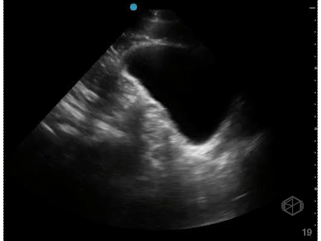
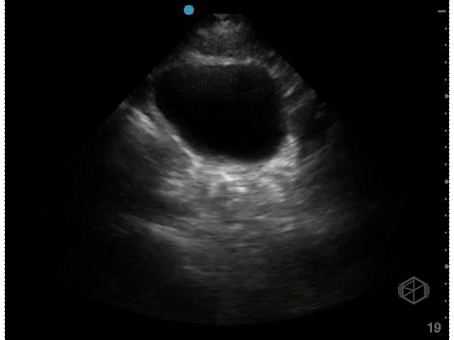
Right kidney
Right kidney easier to visualize.
More anterior and lower than the left kidney due to the liver.
Visualize right kidney in both longitudinal and transverse orientations.
Measure kidney length.
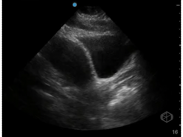
Left kidney
Harder to visualize
More posterior and higher than the right kidney.
Visualize left kidney in both longitudinal and transverse orientations.
Measure kidney length.
May notice “dromedary kidney.”
Bladder views
Include two views of the bladder.
Start sagittal and identify pubic symphysis, fan through entire bladder.
Switch to transverse and fan through entire bladder.
Females — uterus posterior to bladder (unless hysterectomy)
Males — prostate anterior and inferior to bladder; may see prominent seminal vesicles (do not mistake for free fluid).
Limitations:
Empty bladder
Too high gain
Evaluate for hyperechoic density with posterior shadowing (stone); twinkling artifact may be present.
Can evaluate for the presence of ureteral jets although not necessary.