
POCUS Echocardiography
General notes —
There are a few ways to assess LV function: qualitatively and quantitatively.
Qualitative evaluation simply means eye-balling the function, is it: hypodynamic, normal, or hyperdynamic.
This skill comes with time and evaluating multitudes of scans.
Quantitative evaluation includes measurements such as E-point septal separation, fractional shortening, fractional area of change, and Simpson’s biplane method of discs.
Qualitative evaluation —
Look at the overall appearance of the left ventricle in multiple views, especially parasternal short. How well do the walls come together in systole?
There should be a >50% change in diameter if normal EF.
With irregular rhythms need to evaluate average, can be difficult.
Look at the anterior mitral valve leaflet? Is it nearly touching the septum during diastole?
Can be limited by abnormal mitral valves or papillary muscle rupture.
This method can have a high accuracy, especially for normal and low LVEF. Can have a tendency to underestimate LV function.
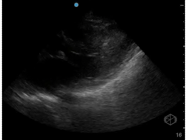
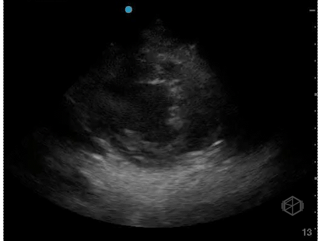
Hyperdynamic LVEF (>70%)
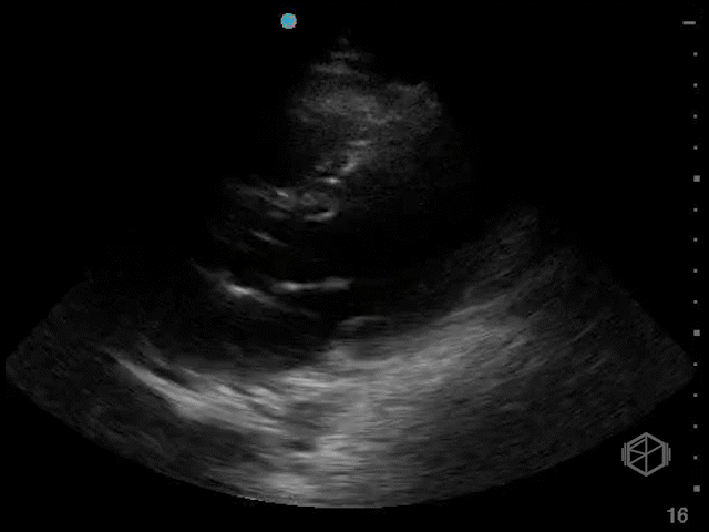
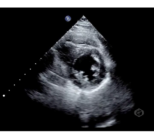
Normal LVEF (~55%)
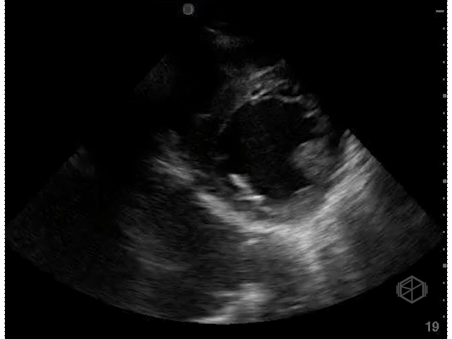
Moderately Hypodynamic LVEF (~35%)
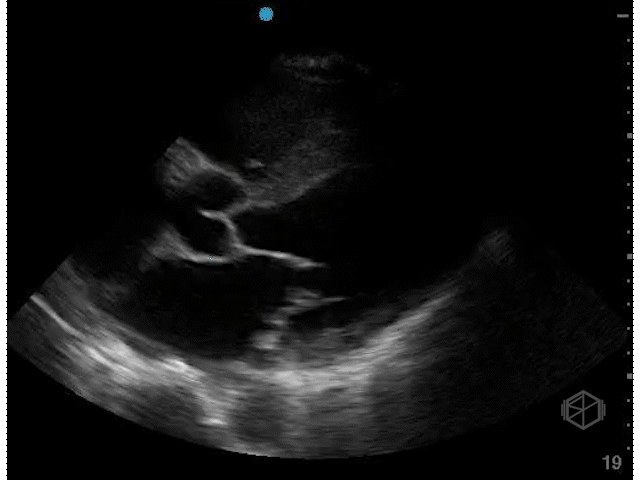
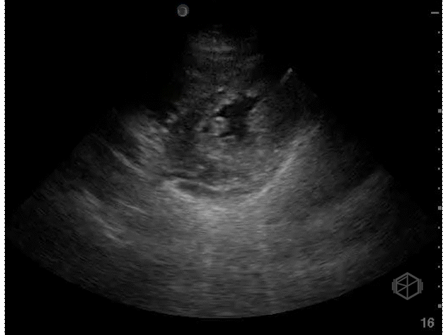
Severely hypodynamic LVEF (~15%)
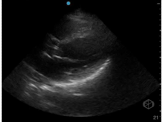
Hyperdynamic LVEF PSS (>70%)
Normal LVEF (~55%)
Moderately hypodynamic LVEF (~30's)
Severely hypodynamic LVEF (~15%)
Quantitative evaluation — E-point septal separation
E-point septal separation is a quantitative method of approximating LV function in the PSL view. It is not a volume assessment.
It can be a reliable method of determining LV function, although with significant limitations.
Some argue that EPSS is not reliable and should not be used compared to qualitative evaluation
(📚 PMID: 34654586).
It looks at how close the anterior leaflet of the mitral valve gets to the septum during early diastolic filling, the E-point.
In a normal heart the E-point should be very close to the septum in early diastole.
EPSS > 7mm indicates reduced EF.
EPSS ≥ 13mm indicates severely reduced EF (<35%) in some references.
Limitations of EPSS:
Abnormalities of valves — mitral valve replacement, calcifications or masses, aortic regurgitation.
Abnormalities of the septum — septal hypertrophy may lead to an artificially reduced EPSS.
Abnormalities of rhythm — irregular rhythms lead to a beat-to-beat variability.
One MRI study proposed this formula to elicit a numerical ejection fraction from EPSS: EF = 75.5-2.5 (EPSS in mm) — although this has limitations.