
POCUS FAST/FAFF
General notes —
Must be in abdominal or eFAST setting.
Must include RUQ, LUQ, and two views of the bladder at the least. Cardiac window recommended but not always necessary dependent on clinical suspicion.
Clips must demonstrate above the diaphragm to below the inferior renal pole, can be done in multiple clips or one clip, with fanning and sliding motions.
Free fluid generally makes sharp angles (V-shapes).
If free fluid detected, continue to obtain other windows as the source may be discovered.
A negative FAST/FAFF does not rule out emergent pathologies. It is a rule IN test, not a rule out test.
Must document follow-up study if negative FAST/FAFF exam — repeat serial abdominal exams or POCUS exams as indicated, if not obtaining cross-sectional imaging.
False negatives, in general, occur due to not fanning through and sliding the probe around to evaluate for free fluid. One single view of each area is not enough to adequately determine if free fluid present.
Extended FAST adds the evaluation for pneumothorax: evaluate for lung sliding and the presence of lung point.
Indications —
In the unstable trauma patient with blunt trauma can help determine the next steps.
In the other patients can be used for abdominal pain, concern for ectopic pregnancy, evaluation of hypotension.
The role of FAST examination in pediatrics is controversial, there is a much lower sensitivity and specificity in pediatrics.
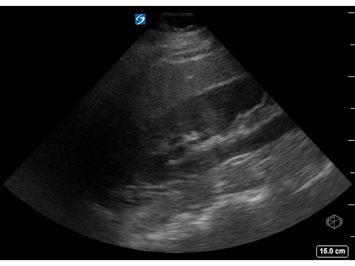
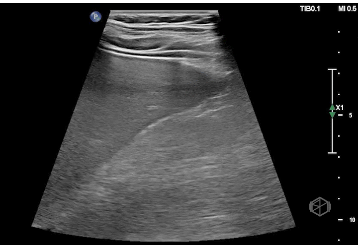
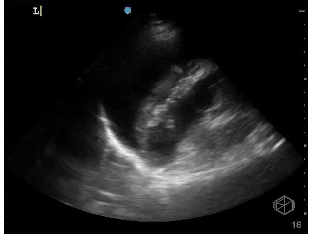
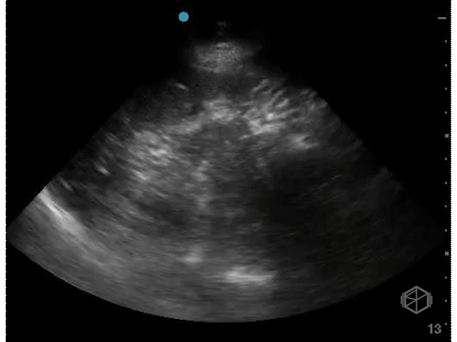
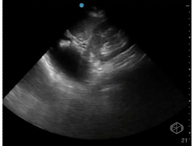
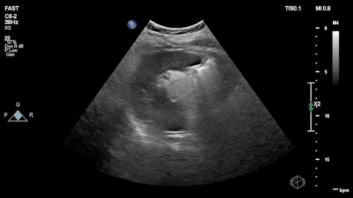
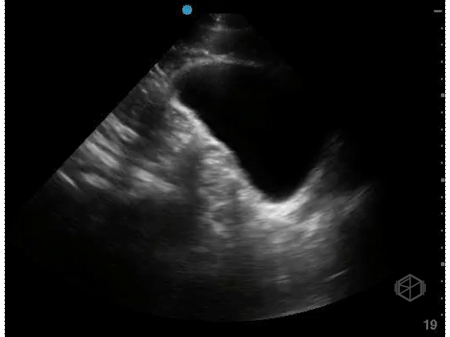
RUQ view —
Include above diaphragm, below diaphragm and between liver, hepatorenal recess, caudal liver edge, and inferior pole of right kidney.
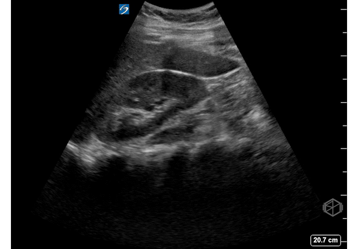
Most sensitive area for free fluid in RUQ = caudal liver edge, not the hepatorenal recess (Morrison’s pouch).
Hepatorenal pouch is negative, however large volume of free fluid noted at the caudal liver edge.
Small volume free fluid at the caudal liver edge.
Positive Hepatorenal (Morrison's Pouch)
RUQ common false positives —
Gallbladder — probe too anterior on body, appears as a walled structure as opposed to sharp angles.
Hepatic veins — use color doppler if suspicious for subtle free fluid versus hepatic veins.
Focal fatty sparing — localized absence of increased intracellular hepatic fat, in a liver otherwise fatty in appearance.
Double line sign — occurs due to perinephric fat.
IVC — probe too posterior, appears with respiratory variation with thin walls.
Renal cysts — rounded, does not make sharp angles, but complex cysts may be mistaken for free fluid.
Lipliner sign — post-processing artifact in modern ultrasound machines that creates a thin, hypoechoic linear outline along the edge of a solid organ (e.g. liver or spleen), which can be mistaken for free fluid.
This artifact arises from “image adaptive” filtering or speckle-reduction algorithms (which smooth the image) rather than true anatomy, and its recognition is important to avoid false positive diagnoses of intra-abdominal bleeding.
Double line sign
Focal fatty sparing
Gallbladder as a false positive
Lipliner sign near liver tip
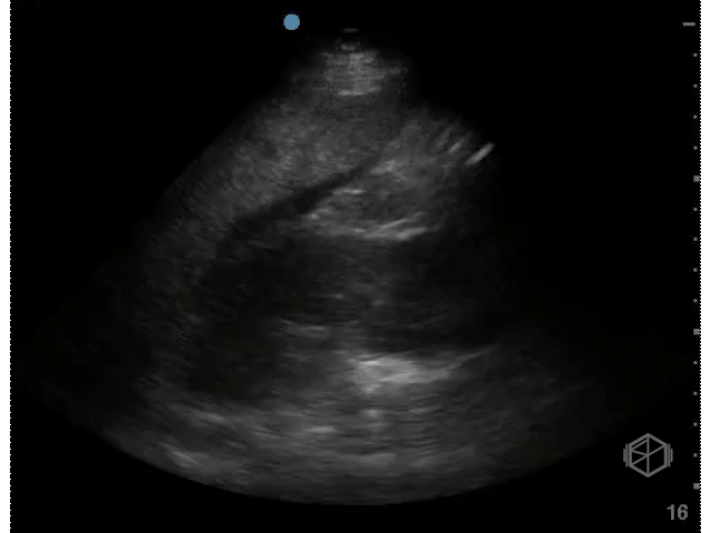
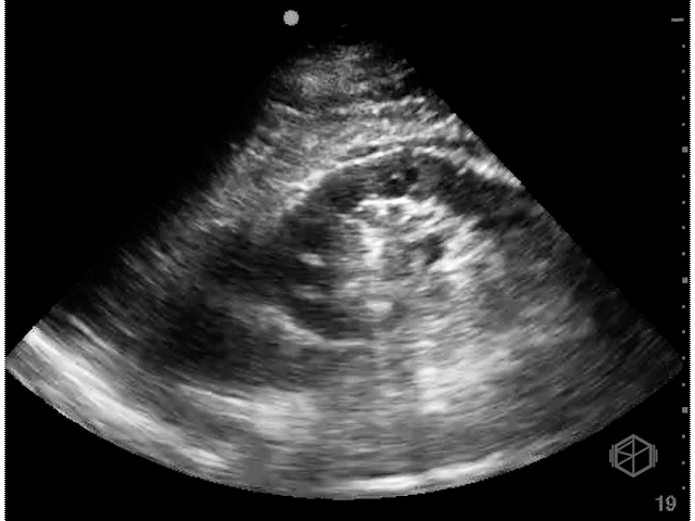
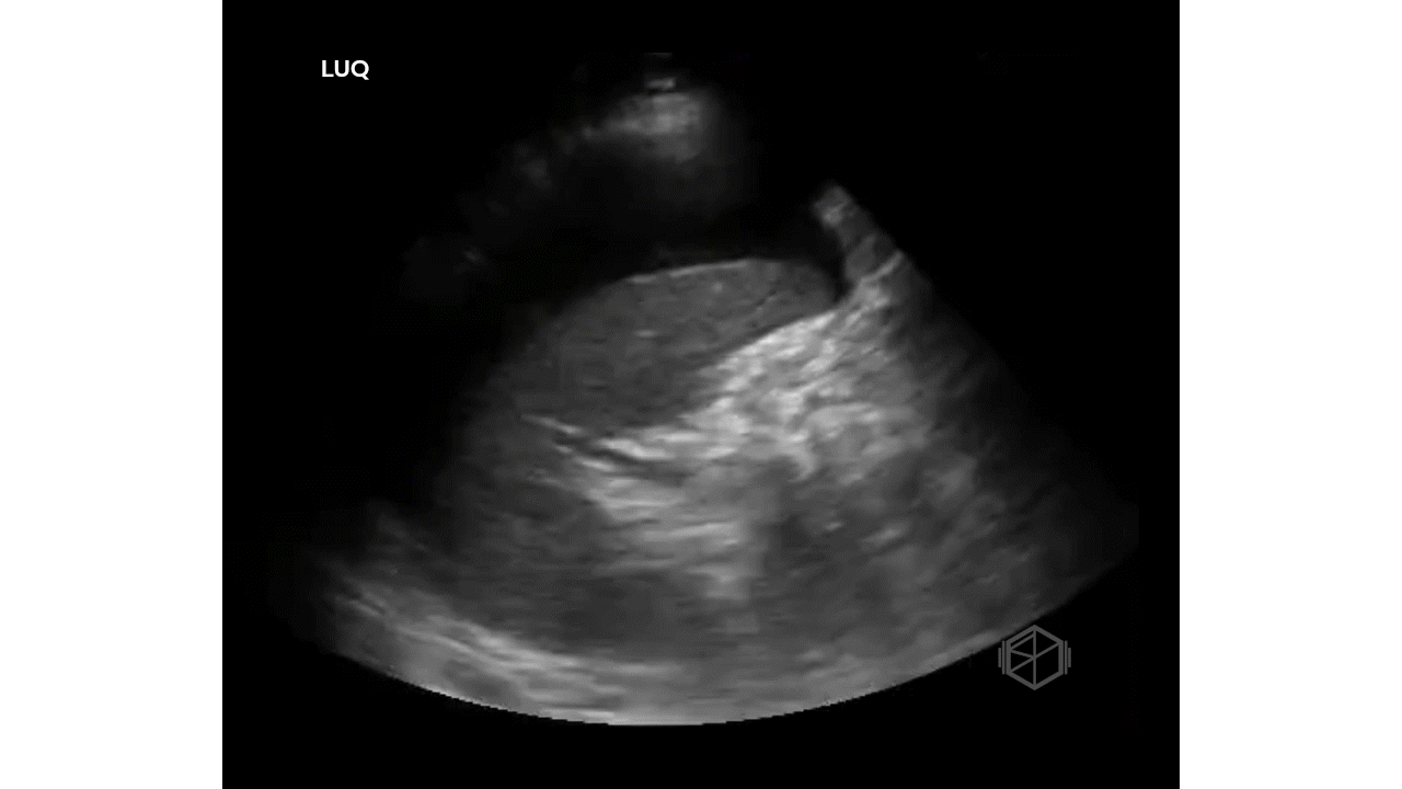
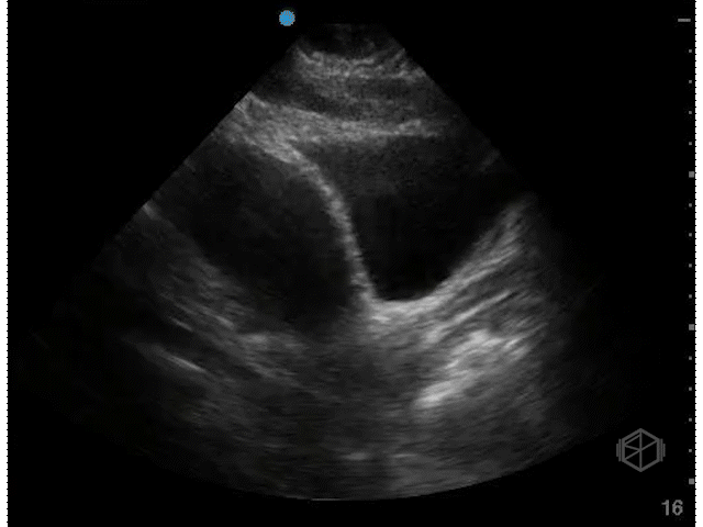
LUQ view —
Include above diaphragm, below diaphragm and between spleen, splenorenal recess, and inferior pole of left kidney.
More difficult view to obtain due to being more posterior and spleen does not provide a good sonographic window.
Most sensitive area for free fluid in LUQ = splenodiaphragmatic space or paracolic gutter.
+LUQ free fluid
+LUQ large volume free fluid
+LUQ splenodiaphragmatic space
LUQ common false positives —
Stomach — probe too anterior, will see curved structure with rugae and perhaps food particles.
Renal cysts — rounded, do not make sharp angles.
Double line sign — occurs due to perinephric fat
Lipliner sign — post-processing artifact in modern ultrasound machines that creates a thin, hypoechoic linear outline along the edge of a solid organ (e.g. liver or spleen), which can be mistaken for free fluid.
This artifact arises from “image adaptive” filtering or speckle-reduction algorithms (which smooth the image) rather than true anatomy, and its recognition is important to avoid false positive diagnoses of intra-abdominal bleeding.
Stomach as false positive in the LUQ.
Lipliner sign with Spleen
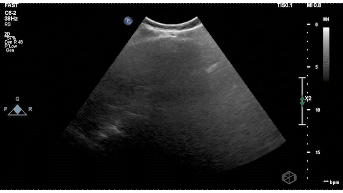
Pelvis view—
Include two views of the pelvis area.
Start sagittal and identify pubic symphysis, fan through entire bladder.
Switch to transverse and fan through entire bladder.
Limitations —
Empty bladder
Too high gain / posterior acoustic enhancement
Common false positives —
Iliac vessels — can be rarely misinterpreted as free fluid. Use color doppler, they lie lateral to the bladder.
Males — prominent seminal vesicles.
Females — physiologic free fluid, fibroids, ovarian cysts.
In generally better to call equivocal and obtain further imaging if high suspicion.
Correlate clinically, free fluid can be physiologic in females. Physiologic free fluid is usually deep to the uterus, if the free fluid extends beyond 1/3rd the border of the uterus, consider pathologic free fluid.
Normal Male Sagittal
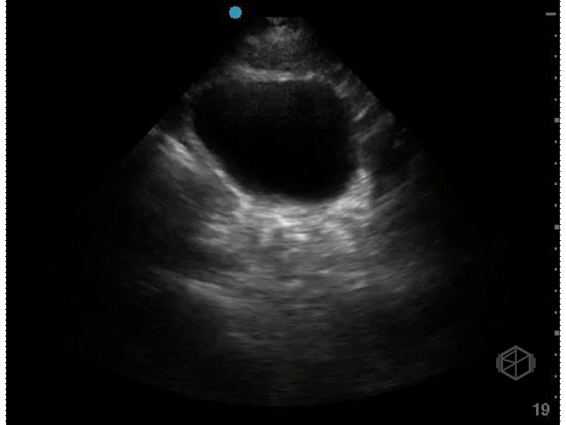
Normal Male Transverse
Normal Female Sagittal
Normal Female Transverse
+Free fluid in female, sagittal view.
+Free fluid in female, transvere view.
+Free fluid, male, note how the high gain makes it difficult to see free fluid.
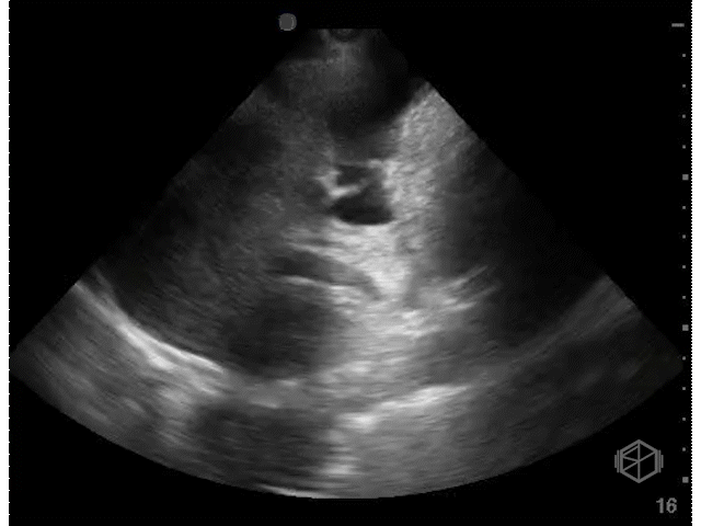
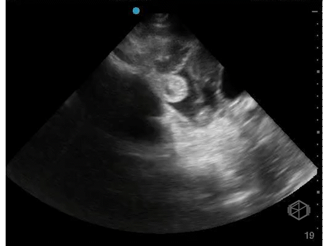
Cardiac —
See echo section on pericardial effusions.
Can use either subxiphoid or parasternal long depending on patient’s habitus or limitations.
When asking the question of free fluid/pericardial effusion, subtle other cardiac findings may be missed, simply write “no pericardial effusion,” rather than “negative study” or “normal study.”
If pericardial effusion is suspected, obtain other views to confirm presence of pericardial effusion.
False negatives —
May miss subtle free fluid when not fanning through the heart.
Common false positive:
Pericardial fat pad —
Pericardial fat pad is echogenic/heterogeneous, move with the heart, and is anterior.
Pericardial effusions are circumferential, can be anechoic or heterogenous depending on simple versus complex, but are usually stationary.